. 2025 Feb 4;16(1):1348.
doi: 10.1038/s41467-025-56610-6.

Molecular basis for azetidine-2-carboxylic acid biosynthesis

Affiliations

Molecular basis for azetidine-2-carboxylic acid biosynthesis

Tim J Klaubert et al. Nat Commun. .

Abstract

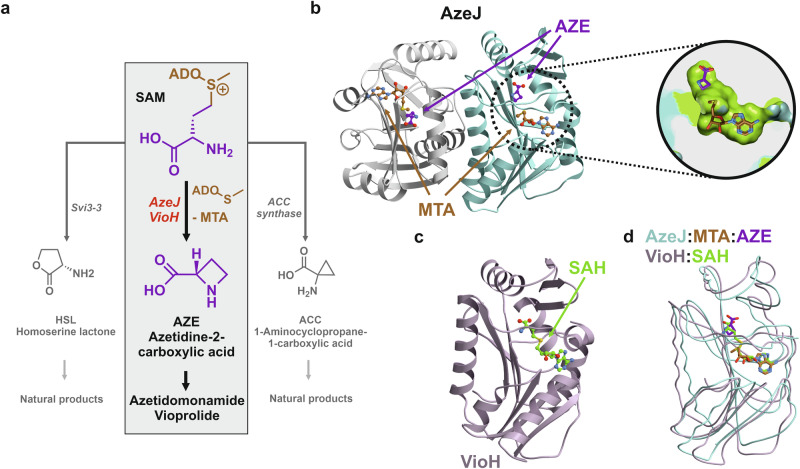
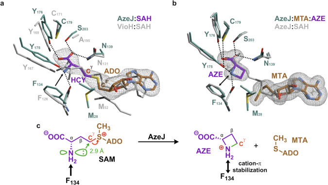
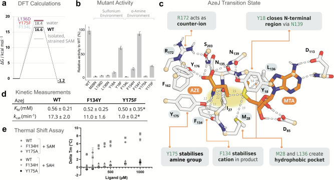
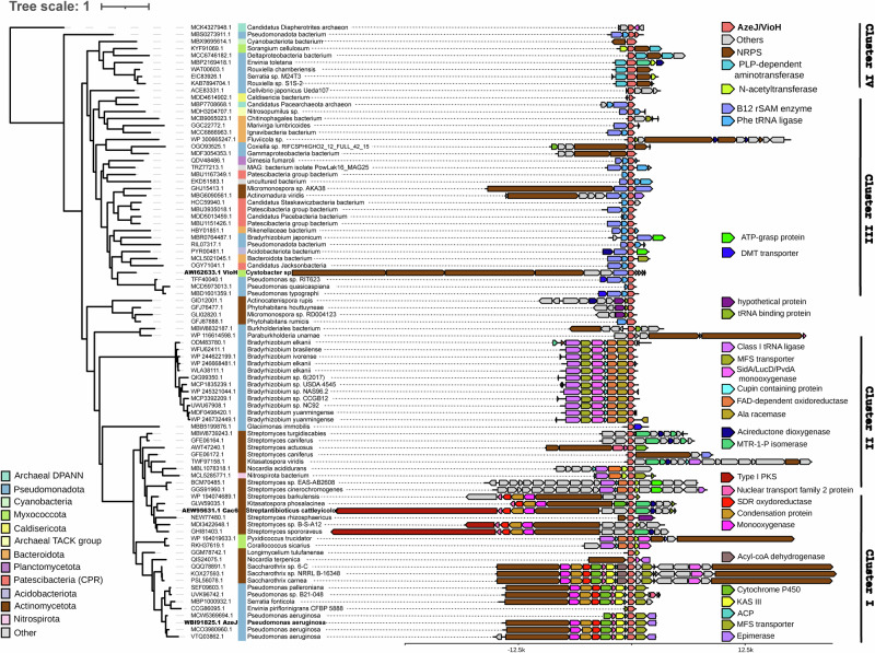
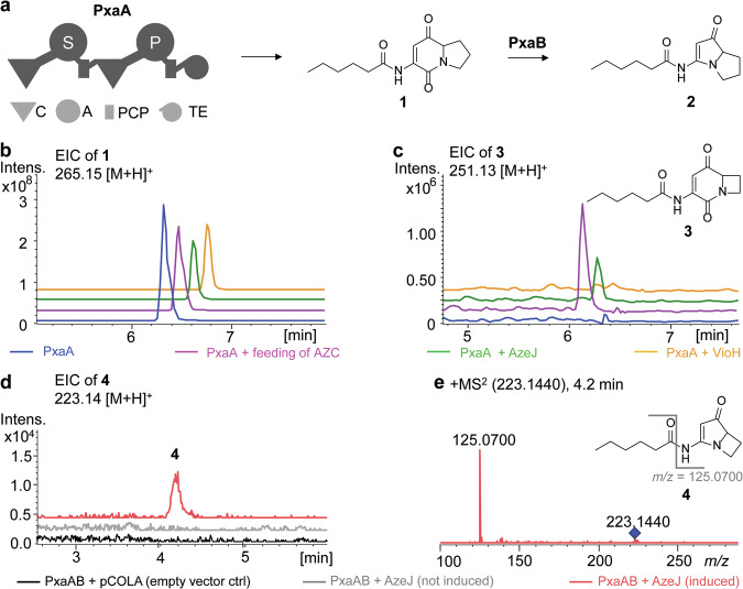
Azetidine-2-carboxylic acid (AZE) is a long-known plant metabolite. Recently, AZE synthases have been identified in bacterial natural product pathways involving non-ribosomal peptide synthetases. AZE synthases catalyse the intramolecular 4-exo-tet cyclisation of S-adenosylmethionine (SAM), yielding a highly strained heterocycle. Here, we combine structural and biochemical analyses with quantum mechanical calculations and mutagenesis studies to reveal catalytic insights into AZE synthases. The cyclisation of SAM is facilitated by an exceptional substrate conformation and supported by desolvation effects as well as cation-π interactions. In addition, we uncover related SAM lyases in diverse bacterial phyla, suggesting a wider prevalence of AZE-containing metabolites than previously expected. To explore the potential of AZE as a proline mimic in combinatorial biosynthesis, we introduce an AZE synthase into the pyrrolizixenamide pathway and thereby engineer analogues of azabicyclenes. Taken together, our findings provide a molecular framework to understand and exploit SAM-dependent cyclisation reactions.


Conflict of interest statement

Competing interests: The authors declare no competing interests.

Figures

Fig. 1
Fig. 1. X-ray structures of AzeJ and VioH.
a Intramolecular cyclisation of S-adenosylmethionine (SAM) can result in three-, four-, or five-membered ring products. Enzymes catalysing 1-aminocyclopropane-1-carboxylic acid (ACC), azetidine-2-carboxylic acid (AZE), and homoserine lactone (HSL) include aminocyclopropane-1-carboxylic acid (ACC) synthase, AzeJ/VioH and Svi3-3, respectively. b Ribbon drawing of the AzeJ homodimer (PDB ID: 8RYE) bound to 5′-methylthioadenosine (MTA, brown) and AZE (purple). The close-up provides a cut open view of the active site surface (carbons in green) in complex with MTA and AZE (sticks). c Ribbon drawing of monomeric VioH (PDB ID: 8RYG) bound to S-adenosyl-L-homocysteine (SAH, green). d The overlay of AzeJ:MTA:AZE (turquoise) and VioH:SAH complex structures (light purple) indicates their structural similarity.
Fig. 2
Fig. 2. Molecular insights into the active sites of AzeJ and VioH.
a Close-up of the catalytic centre of AzeJ:SAH (PDB ID: 8RYD), highlighting the specificity pocket (residues in cyan, adenosyl residue (ADO) in brown, homocysteine (HCY) in purple). The VioH:SAH complex structure is superimposed (PDB ID: 8RYG, C-atoms in grey). Residues are numbered according to the primary sequence of the respective enzyme. In AzeJ, the activation of the NH2 group of HCY via F134 π-interactions is shown by a black arrow. b Crystallisation of AzeJ in the presence of SAM resulted in the AzeJ:MTA:AZE complex (PDB ID: 8RYE, MTA in brown, AZE in purple). The amine group of AZE forms hydrogen bonds with the hydroxyl group of Tyr175 and the carbonyl oxygen of Phe134. Strong cation-π interactions with Phe134 (black double arrow) indicate a protonated state of the nitrogen atom. The overlay with AzeJ:SAH (grey) illustrates catalysis without structural rearrangements. The 2Fo-Fc electron density maps (1σ, grey mesh) are displayed for ligands that were omitted for phasing. c Proposed catalytic mechanism of AzeJ. Left panel: The electrophilic Cγ-carbon (red) is attacked by the amine (distance 2.9 Å). Right panel: The positive charge is transferred from SAM to the AZE nitrogen atom and stabilised by cation-π interactions with Phe134 (black double arrow).
Fig. 3
Fig. 3. Computational and biochemical characterisation support the proposed mechanism of AzeJ catalysis.
a Electronic energies of reactant (SAM), transition and product states (AZE + MTA) for AzeJ wild-type (WT) and mutants based on DFT cluster models of the active site (Fig. S12), calculated at the B3LYP-D3/def2-TZVP/ε = 4 level. Comparative reactions in solvent were modelled using implicit solvation (in water, ε = 80; hydrophobic, ε = 4). More details are provided in the Methods section and Table S5. b Relative activity of AzeJ variants compared to WT. Data are presented as mean values ± standard deviation (SD) with three replicates. Experiments are repeated twice independently. c DFT model of the AzeJ WT transition state (alternative view in Fig. S16). Highlighted residues were analysed by mutagenesis; atoms shown in light orange were fixed during geometry and reaction pathway optimisations. For clarity, only polar hydrogens are shown (distances in Å). d Determination of kinetic parameters (KM, kcat, *denotes apparent parameters without consideration of substrate inhibition) (graphs see Fig. S19). e Evaluation of ligand binding by thermal shift assays for AzeJ WT, F134Y and Y175F variants. Data are presented as mean values ± SD with two biological replicates. Source data are provided as a Source Data file for (b, d and e).
Fig. 4
Fig. 4. Maximum likelihood phylogenetic tree and genomic context analysis of AZE synthases.
Accession numbers in bold highlight AzeJ, VioH and Cac6, involved in the biosynthesis of azetidomonamides, vioprolides and clipibicyclene, respectively. The colour code illustrates relevant neighbouring genes encoding AzeJ-related products (coral pink), NRPSs (brown), B12-dependent rSAM enzymes (purple), class I tRNA-ligase (magenta), ATP-grasp enzymes (bright green), N-acetyltransferase (apple green) and drug/metabolite (DMT) transporter (cobalt). Source data are provided as a Source Data file.
Fig. 5
Fig. 5. Production of AZE-incorporated pyrrolizixenamides.
a Native biosynthetic scheme of pyrrolizixenamides involving the dimodular nonribosomal peptide synthetase PxaA together with the Baeyer-Villiger monooxygenase PxaB. Domains: C (condensation), A (adenylation), PCP (peptidyl carrier protein), TE (thioesterase). Domain specificity is indicated in white. b, c Extracted ion chromatograms (EICs) of indicated compounds under various conditions: expression of pxaA in E. coli (blue), feeding AZE to a pxaA-expressing E. coli strain (pink), co-expression of pxaA and azeJ (green), as well as co-expression of pxaA and vioH (yellow) in E. coli. LC-HRMS analysis confirmed the identity of 1 (m/z calc’d for C14H20N2O3 [M + H]+ = 265.1546; obs’d. m/z = 265.1548; Δppm = 0.3) and 3 (m/z calc’d for C13H18N2O3 [M + H]+ = 251.1390; obs’d. m/z = 251.1390; Δppm = 0) (Fig. S23). Production of the azetidomonamide B (azabicyclene) analogue 4 upon co-expressing pxaAB and azeJ in E. coli: empty vector control (black), uninduced (grey), induced (red). The identity of 4 was confirmed by EIC (d) and by tandem mass spectrometry (e). Inset: proposed structure of 4 (m/z calc’d for C12H18N2O2 [M + H]+ = 223.1441; obs’d. m/z = 223.1440; Δppm = −0.6). The key MS2 fragment is indicated (m/z calc’d for C6H8N2O [M + H]+ = 125.0709; obs’d. m/z = 125.0700; Δppm = −7.2).

References

    1. Lee, Y.-H., Ren, D., Jeon, B. & Liu, H. S-Adenosylmethionine: more than just a methyl donor. Nat. Prod. Rep.40, 1521–1549 (2023). - PMC - PubMed
    1. Sun, Q., Huang, M. & Wei, Y. Diversity of the reaction mechanisms of SAM-dependent enzymes. Acta Pharm. Sin. B11, 632–650 (2021). - PMC - PubMed
    1. Cornforth, J. W., Redmond, J. W., Eggerer, H., Buckel, W. & Gutschow, C. Asymmetric methyl groups: asymmetric methyl groups, and the mechanism of malate synthase. Nature221, 1212–1213 (1969). - PubMed
    1. Cornforth, J. W., Redmond, J. W., Eggerer, H., Buckel, W. & Gutschow, C. Synthesis and configurational assay of asymmetric methyl groups. Eur. J. Biochem.14, 1–13 (1970). - PubMed
    1. Guo, X. et al. Structure and mechanism of a phage-encoded SAM lyase revises catalytic function of enzyme family. eLife10, e61818 (2021). - PMC - PubMed

MeSH terms

LinkOut - more resources