. 2025 Feb 4;31(1):45.
doi: 10.1186/s10020-025-01090-z.

Rescue of common and rare exon 2 skipping variants of the GAA gene using modified U1 snRNA

Affiliations

Rescue of common and rare exon 2 skipping variants of the GAA gene using modified U1 snRNA

Paolo Peruzzo et al. Mol Med. .

Abstract

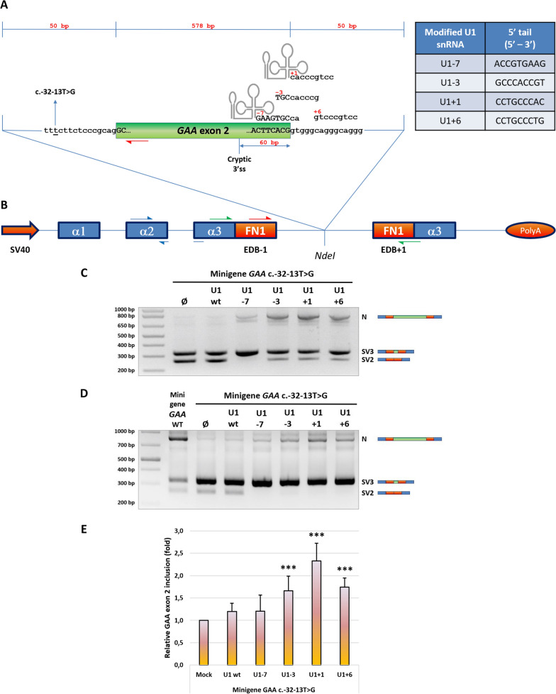
Background: Pompe disease (PD) is an autosomal recessive lysosomal storage disorder caused by the deficient activity of acid alpha glucosidase (GAA) enzyme due to mutations in the GAA gene. As a result, undigested glycogen accumulates within lysosomes causing their dysfunction. From a clinical point of view, the disease can be classified in infantile-onset (IO) and late-onset (LO) forms. The common GAA c.-32-13T>G variant, found in 40-70% of LO-PD alleles, is a leaky splicing mutation interfering with the correct GAA exon 2 recognition by the spliceosome leading to the production of non-functional GAA transcripts. In this study, we used modified, GAA-tailored U1 snRNAs to correct the aberrant splicing determined by the c.-32-13T>G and other GAA exon 2-skipping mutations.

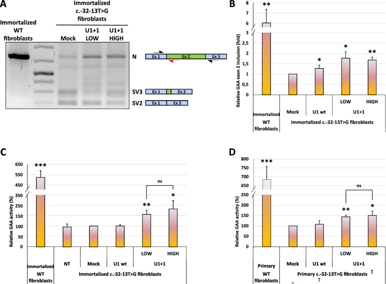
Methods: A set of constructs expressing 5 different engineered U1 snRNAs was generated. A functional splicing assay using a GAA hybrid minigene carrying different variants known to affect GAA exon 2 splicing was used to test the effect of engineered U1 snRNAs on exon 2 inclusion. The effect on endogenously expressed GAA transcript and GAA enzymatic activity was assessed by transfecting patient-derived fibroblasts bearing the common c.-32-13T>G with the best performing modified U1 snRNA.

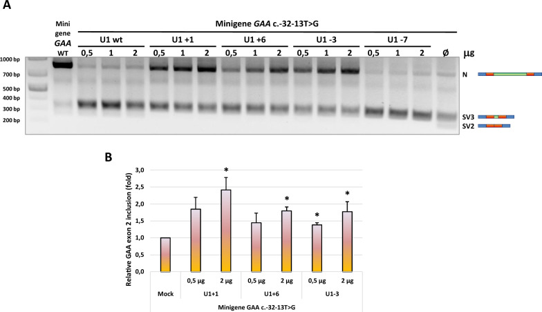
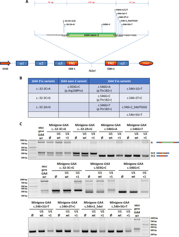
Results: Modified U1-3, U1+1 and U1+6 snRNAs were all able to increase, in a dose-dependent manner, the inclusion of exon 2 within the transcript derived from the GAA minigene harbouring the c.-32-13T>G variant. The U1+1 was the most effective one (2,5 fold increase). Moreover, U1+1 snRNA partially rescued the correct splicing of GAA minigenes harbouring mutations that affect the 3'ss (c.-32-3C>G, c.-32-2A>G) and the 5'ss (c.546G>A, c.546G>C, c.546G>T). Notably, the treatment of patient-derived fibroblasts carrying the c.-32-13T>G mutation with the U1+1 snRNA increased the amount of normal GAA mRNA by 1,8 fold and the GAA enzymatic activity by 70%.

Conclusions: we provide the proof-of-concept for the use of modified GAA-tailored U1 snRNAs, designed to potentiate the recognition of the GAA exon 2 5'ss, as therapeutic tools to correct the aberrant transcripts carrying variants that affect exon 2 splicing, including the common c.-32-13T>G variant.

Keywords: GAA; Glycogenosis type II; Pompe disease; Splicing; U1; c.-32-13T>G.


Conflict of interest statement

Declarations. Ethics approval and consent to participate: Not applicable. Consent for publication: Not applicable. Competing interests: The authors have no conflict of interests to declare.

Figures

Fig. 1
Fig. 1
Effect of modified U1 snRNAs −7, −3, +1 and +6 on the splicing of GAA c.-32-13T>G (MUT) minigene. A The schematic diagram shows the GAA minigene sequences targeted by the modified U1 snRNAs. The table reports, for each modified U1 snRNA, also the specific 9 bp-long tail sequence (5’ to 3’) that ensures complementarity to the selected positions within GAA intron 2 donor splice site. B Schematic representation of the human GAA minigene system: the entire GAA exon 2 (green box) and 50 nucleotides of both flanking introns were cloned within the NdeI restriction site in the splicing-proficient pTB vector containing the Alfa Globin (blue boxes) and fibronectin extra-domain B (EDB - red boxes) exons. The MUT minigene harbouring the GAA c.-32-13T>G variant (underscored) was obtained by site-directed mutagenesis from the GAA WT minigene. The position of the cryptic 3’ splice site 60 bp upstream the end of GAA exon 2 is highlighted. Primers Alfa 2–3 FOR and Bra 2 REV used for the minigene functional splicing assay are represented by green half-arrows, whereas primer couples GAA mini FOR/GAA mini REV and Alfa Glo FOR/Alfa Glo REV used for the quantification of GAA exon 2 inclusion by real-time qPCR are depicted by red and blue half-arrows, respectively. C and D Functional splicing assay of MUT minigene after modified U1 snRNAs co-transfection performed on HeLa and Hek 293 cell lines, respectively. The RT-PCR amplified bands correspond to the normal, GAA exon 2-included, splicing isoform (N) and the aberrant splicing isoforms with complete (SV2; r.-32_546del) or partial (SV3; r.-32_486del) exclusion of GAA exon 2, the latter deriving from the activation of the 3’ cryptic splice site. GAA WT minigene was used as internal control. Agarose gel pictures show a representative result of five independent experiments (E) Real-time qPCR analysis of the extent of GAA exon 2 inclusion (N isoform) in MUT minigene/modified U1 snRNAs co-transfected Hek 293 cells. Data, expressed as fold of inclusion relative to the MUT minigene alone, are presented as mean ± SD of four independent experiments. Statistical analysis was conducted using Student t-test. *** = p-value < 0,001
Fig. 2
Fig. 2
Effect of dose-escalation of modified U1 snRNAs −7, −3, +1 and +6 on the splicing of GAA c.-32-13T>G (MUT) minigene. A Functional splicing assay performed on Hek 293 cells co-transfected with 0,5 µg of MUT minigene and increasing doses (0,5 – 1 – 2 µg) of the different modified U1 snRNAs. The minigene-specific N, SV3 and SV2 splicing isoforms are evidenced on the right. The agarose gel picture shows a representative result of two independent experiments. B Real-time qPCR analysis of the extent of GAA exon 2 inclusion (N isoform) in MUT minigene/effective modified U1 snRNAs co-transfected Hek 293 cells. Data, expressed as fold of inclusion relative to the MUT minigene alone, are presented as mean ± SD of two independent experiments. Statistical analysis was conducted using Student t-test. * = p-value < 0,05
Fig. 3
Fig. 3
Effect of modified U1+1 snRNA on the splicing of mutant GAA minigenes harbouring the 11 selected GAA splicing-affecting variants. A The cartoon indicates the position of the 11 selected GAA splicing variants within the GAA insert of the minigene plasmid. All the GAA mutant minigenes were obtained by site-directed mutagenesis from the GAA WT minigene (Goina E et al., 2019). (B) The table reports the distribution of the 11 selected GAA splicing variants: three of them (c.-32-3C>A, c.-32-3C>G and c.-32-2A>G) fall within the 3’ splice site; one (c.503G>C) is exonic and seven (c.546G>A, c.546G>C, c.546G>T, c.546+1G>T, c.546+2T>C, c.546+2_5delTGGG and c.546+5G>T) involve the 5’ splice site. C Functional splicing assay performed on Hek 293 cells co-transfected with 0,5 µg of the reported GAA mutant minigene and 1,5 µg of wild-type U1 or modified U1+1 snRNAs. For each set, the transfection with the minigene alone was considered the reference sample. GAA WT minigene was used as internal control in each agarose gel. The minigene-specific N, SV3 and SV2 splicing isoforms are evidenced on the right
Fig. 4
Fig. 4
Effect of modified U1+1 snRNA on the splicing of endogenous GAA in immortalized and primary c.-32-13T>G fibroblasts. Functional splicing assay performed on endogenous GAA mRNA in patient-derived immortalized fibroblasts carrying the c.-32-13T>G variant transfected with 2 doses (LOW = 5,8 g; HIGH = 11,6 g) of modified U1+1 snRNA plasmid. The mock-transfected fibroblasts were considered as the reference sample. The GAA N, SV3 and SV2 splicing isoforms and the position of the primers GAA WT FOR and GAA SKIP2 REV (black half-arrows) are evidenced on the right. The red half-arrow represents the GAA WT REV primer used, in combination with the GAA WT FOR primer, to quantify the endogenous GAA N isoform in real-time qPCR experiments. (B) Real-time qPCR analysis of the extent of GAA exon 2 inclusion (N isoform) in c.-32-13T>G fibroblasts transfected with modified U1+1 snRNAs at low and high doses. The transfection with U1 wt was used as negative snRNA control condition. The data, normalized to the expression of HPRT house-keeping gene and displayed as fold of inclusion relative to the mock-transfected c.-32-13T>G fibroblasts, are presented as mean ± SD of three independent experiments. Statistical analysis was conducted using Student t-test. * = p-value < 0,05; ** = p-value < 0,01. C GAA enzymatic activity measured in immortalized c.-32-13T>G fibroblasts transfected with U1+1 snRNA at low and high doses. The transfection with U1 wt was used as negative snRNA control condition. Data, expressed as percentage increase relative to the mock-transfected c.-32-13T>G fibroblasts, are presented as mean ± SD of three independent experiments. Statistical analysis was conducted using Student t-test. * = p-value < 0,05; ** = p-value < 0,01; *** = p-value < 0,001; ns = not significant. D GAA enzymatic activity measured in primary c.-32-13T>G fibroblasts electroporated with U1+1 snRNA at low and high doses. The electroporation with U1 wt was used as negative snRNA control condition. Data, expressed as percentage increase relative to the mock c.-32-13T>G fibroblasts, are presented as mean ± SD of three independent experiments. Statistical analysis was conducted using Student t-test. * = p-value < 0,05; ** = p-value < 0,01; *** = p-value < 0,001; ns = not significant. Throughout the figure, the values obtained in wild type immortalized (panel A, B and C) and primary (panel D) fibroblasts were reported for comparison

References

    1. Balestra D, Scalet D, Ferrarese M, Lombardi S, Ziliotto N, Croes CC, Petersen N, Bosma P, Riccardi F, Pagani F, et al. A compensatory U1snRNA partially rescues FAH splicing and protein expression in a splicing-defective mouse model of tyrosinemia type I. Int J Mol Sci. 2020;21(6):2136. 10.3390/ijms21062136. - PMC - PubMed
    1. Baserga SJ, Gesteland SJA, Atkins RF. The RNA World. In: Cold Spring harbor laboratory press. NY; 1993.
    1. Bellotti AS, Andreoli L, Ronchi D, Bresolin N, Comi GP, Corti S. Molecular approaches for the treatment of pompe disease. Mol Neurobiol. 2020;57(2):1259–80. 10.1007/s12035-019-01820-5. - PubMed
    1. Bergsma AJ, Int Groen SLM, van den Dorpel JJA, van den Hout HJMP, van der Beek NAME, Schoser B, Toscano A, Musumeci O, Bembi B, Dardis A, et al. A genetic modifier of symptom onset in Pompe disease. EBioMedicine. 2019;2019(43):553–61. 10.1016/j.ebiom.2019.03.048. - PMC - PubMed
    1. Boerkoel CF, Exelbert R, Nicastri C, Nichols RC, Miller FW, Plotz PH, Raben N. Leaky splicing mutation in the acid maltase gene is associated with delayed onset of glycogenosis type II. Am J Hum Genet. 1995;56(4):887–97. - PMC - PubMed

LinkOut - more resources