

The Topographic Relationships and Geographic Distribution of Prevascular Vitreous Fissures and Cisterns Assessed by Ultrawidefield En Face Vitreous Images
- PMID: 39906410
- PMCID: PMC11791428
- DOI: 10.1016/j.xops.2024.100660
The Topographic Relationships and Geographic Distribution of Prevascular Vitreous Fissures and Cisterns Assessed by Ultrawidefield En Face Vitreous Images
Abstract
Purpose: To determine the topographic relationships and geographic distribution of prevascular vitreous fissures (PVFs) and cisterns across the entire posterior vitreous membrane in healthy subjects, using ultrawidefield en face and cross-sectional swept-source OCT (SS-OCT) images.
Design: Observational cross-sectional study.
Participants: Ninety-six eyes of 96 healthy participants (age range, 20-49 years) without posterior vitreous detachment.
Methods: For each eye, a 29 × 24-mm SS-OCT volume scan was obtained, along with standardized horizontal and vertical scans through the fovea.
Main outcome measures: Ultrawidefield en face and cross-sectional images were analyzed to assess the topographic relationships and geographic distribution of PVFs and cisterns in the posterior vitreous.
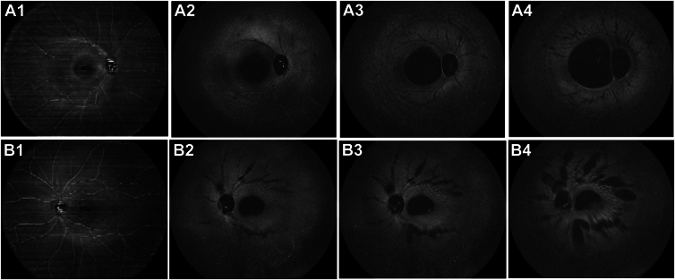
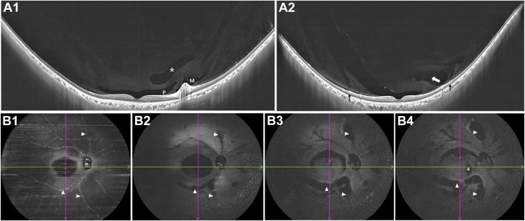
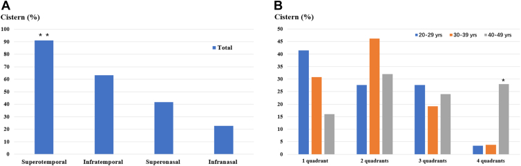
Results: En face imaging readily distinguished various preretinal liquefaction spaces throughout the posterior vitreous, extending to near the equator. Aside from the posterior precortical vitreous pocket (PPVP) and the area of Martegiani, all preretinal liquefied fissures and cisterns were distributed along superficial retinal vessels, suggesting they originated from prevascular vitreous liquefaction. In 96 eyes of healthy young and middle-aged adults, PVFs were identified in all participants, presenting a continuous course. Cisterns were detected in 79 eyes (82.3%) and were distributed as follows: superotemporal (91.1%), infratemporal (63.3%), supranasal (41.8%), and inferonasal (22.8%), respectively. The superotemporal cistern was most frequently observed (P < 0.001), and cisterns were more likely to involve multiple quadrants with age (P = 0.005). Additionally, all preretinal liquefaction spaces, including the PPVP, PVFs, and cisterns, were consistently located overlying the vitreoretinal tightly adhered regions.
Conclusions: Ultrawidefield en face vitreous imaging in healthy young and middle-aged adults revealed that (1) PVFs distributed along superficial retinal vessels with continuous course; (2) cisterns may develop from PVFs and are more common in the superotemporal quadrant; (3) cisterns appear early in life and become more widespread with age; (4) preretinal vitreous liquefaction follows a stereotypic pattern, aligning along regions of firm vitreoretinal adhesion.
Financial disclosures: Proprietary or commercial disclosure may be found in the Footnotes and Disclosures at the end of this article.
Keywords: Cisterns; En face imaging; Prevascular vitreous fissures; Ultra-widefield; Vitreous.
© 2024 by the American Academy of Ophthalmologyé.
Figures
References
-
- Tozer K.J.M., Sebag J. Springer; New York, NY: 2014. Vitreous Aging and Posterior Vitreous Detachment. The Vitreous in Health and Disease. 2014; pp. 131–150.
-
- Worst J.G. Cisternal systems of the fully developed vitreous body in the young adult. Trans Ophthalmol Soc U K (1962) 1977;97:550–554. - PubMed
-
- Kishi S., Shimizu K. Posterior precortical vitreous pocket. Arch Ophthalmol. 1990;108:979–982. - PubMed
-
- Jongebloed W.L., Worst J.F. The cisternal anatomy of the vitreous body. Doc Ophthalmol. 1987;67:183–196. - PubMed
-
- Eisner G. Biomicroscopy of the peripheral fundus. Surv Ophthalmol. 1972;17:1–28. - PubMed
LinkOut - more resources
Full Text Sources
Research Materials
