. 2025 Jan 31;18(1):2458872.
doi: 10.1080/19420889.2025.2458872. eCollection 2025.

Structure simulation-based comparison of active site variations in fungal ornithine decarboxylases

Affiliations

Structure simulation-based comparison of active site variations in fungal ornithine decarboxylases

Min Jeong Kim et al. Commun Integr Biol. .

Abstract

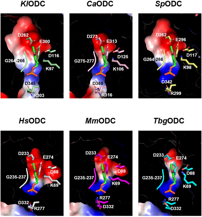
Polyamines play crucial roles in various biological processes, including cell proliferation and differentiation, immune response modulation, and signal transduction. Ornithine decarboxylase (ODC) initiates polyamine biosynthesis by catalyzing the conversion of ornithine to putrescine in a pyridoxal phosphate (PLP)-dependent manner. While the structures of mammalian and protozoan ODCs have been elucidated, fungal ODCs remain uncharacterized. In this study, AlphaFold2 was employed to simulate the structures of ODCs from four fungi: Kluyveromyces lactis, Candida albicans, Debaryomyces hansenii, and Schizosaccharomyces pombe. The results indicated that, although these ODCs share α/β-barrel and β-sheet domains, their active site conformations exhibit subtle differences. Additionally, substrate selectivity among ODCs and related decarboxylases varied depending on the distance between the Cα of aspartate or glutamate residues within the specificity helix and the C4α of PLP. Notably, the bacterial Campylobacter jejuni decarboxylase (CjCANSDC), which binds the largest substrate, exhibits the longest distance, whereas fungal ODC, which binds the smallest substrate, displays the shortest distance. Furthermore, significant differences in the composition of amino acid residues within the active sites were also observed. This study provides insights into the structural diversity and catalytic activity of ODCs across a broad range of organisms, advancing the understanding of structure-dependent evolutionary processes.

Keywords: Candida albicans; Debaryomyces hansenii; Kluyveromyces lactis; PLP-dependent enzyme; Schizosaccharomyces pombe; ornithine decarboxylase; polyamine; specificity helix.


Conflict of interest statement

No potential conflict of interest was reported by the author(s).

Figures

Figure 1.
Figure 1.
The predicted three-dimensional structures of the KlODC, DhODC, CaODC, and SpODC proteins. The structures of the ODCs from four different fungi Kluyveromyces lactis (KlODC), Debaryomyces hansenii (DhODC), Candida albicans (CaODC), and schizosaccharomyces pombe (SpODC) were predicted using AlphaFold2 and visualized using PyMOL. While the number of β-strands was consistent, variations were observed in the number of α-helixes. The differing regions are highlighted using red dotted circles. The right panel shows a 90º rotated view of the N-terminal region of the structures.
Figure 2.
Figure 2.
Comparison of the homologous structures of ODCs. (a) Amino acid sequence alignment of ODCs from various organisms. ODC from Kluyveromyces lactis (kl), Debaryomyces hansenii (dh), Candida albicans (ca), schizosaccharomyces pombe (sp), Homo sapiens (hs), Mus musculus (mm) and Trypanosoma brucei gambiense (tbg) were compared. Among the six species, conserved residues with 100% identity are emphasized in red, and those with 75% identity are denoted by blue boxes. Small and big black dots above the sequences indicate every 10th- and 50th- residues, respectively. Conserved residues with 100% identity are highlighted in red, and those with 75% identity are marked using blue boxes. Black dots above the sequences indicate every 10th residue. (b) The structures of their homologs, HsODC (PDB ID: 1D7K), MmODC (PDB ID: 7ODC), and TbgODC (PDB ID: 1NJJ), were obtained from PDB. (c) The structures of KlODC, DhODC, CaODC, and SpODC were predicted using AlphaFold2. The three-dimensional structures of KlODC, DhODC, CaODC, and SpODC are compared with those of HsODC, MmODC, and TbgODC. Unique α-helices or β-strands specific to each structure are highlighted using red dotted circles, and regions with differing lengths or shapes are highlighted using blue dotted circles.
Figure 3.
Figure 3.
Discrepancy in electrostatic distribution and conformation at the plp-binding pocket. The plp-binding pockets of KlODC, CaODC, SpODC, HsODC, MmODC, and TbgODC. The red and blue surfaces represent acidic and basic residues, respectively.
Figure 4.
Figure 4.
Comparison of the specificity helix sequences and substrate selectivity for CjCANSDC, PBCVADC, and fungal ODCs. (a) The substrates are listed in descending order of size: carboxynorspermidine for CjCANSDC, L-arginine for PBCVADC, and L-ornithine for ODC. (b) The dimer structures of CjCANSDC (PDB ID: 3N29), PBCVADC (PDB ID: 2NVA), and HsODC (PDB ID: 1D7K) are shown with an enlarged view of their PLP and substrate binding sites. The secondary structures and sequences of the specificity helices are shown. In the case of HsODC, the sequences of MmODC and TbgODC are included.

Similar articles

References

    1. Hussain T, Tan B, Ren W, et al. Exploring polyamines: functions in embryo/fetal development. Anim Nutr. 2017. Mar;3(1):7–10. doi: 10.1016/j.aninu.2016.12.002 - DOI - PMC - PubMed
    1. Lian J, Liang Y, Zhang H, et al. The role of polyamine metabolism in remodeling immune responses and blocking therapy within the tumor immune microenvironment. Front Immunol. 2022;13:912279. doi: 10.3389/fimmu.2022.912279 - DOI - PMC - PubMed
    1. Murray-Stewart TR, Woster PM, Casero RA Jr.. Targeting polyamine metabolism for cancer therapy and prevention. Biochem J. 2016. Oct 1;473(19):2937–2953. doi: 10.1042/BCJ20160383 - DOI - PMC - PubMed
    1. Handa AK, Fatima T, Mattoo AK.. Polyamines: Bio-molecules with diverse functions in plant and human health and disease. Front Chem. 2018;6:10. doi: 10.3389/fchem.2018.00010 - DOI - PMC - PubMed
    1. Pegg AE, Casero RA Jr.. Current status of the polyamine research field. Methods Mol Biol. 2011;720:3–35. - PMC - PubMed

LinkOut - more resources