

Anti-PL-7/PL-12 antisynthetase syndrome associated with interstitial lung disease following SARS-COV-2 infection and vaccination: A case study review
- PMID: 39906838
- PMCID: PMC11791273
- DOI: 10.1016/j.heliyon.2024.e41311
Anti-PL-7/PL-12 antisynthetase syndrome associated with interstitial lung disease following SARS-COV-2 infection and vaccination: A case study review
Abstract
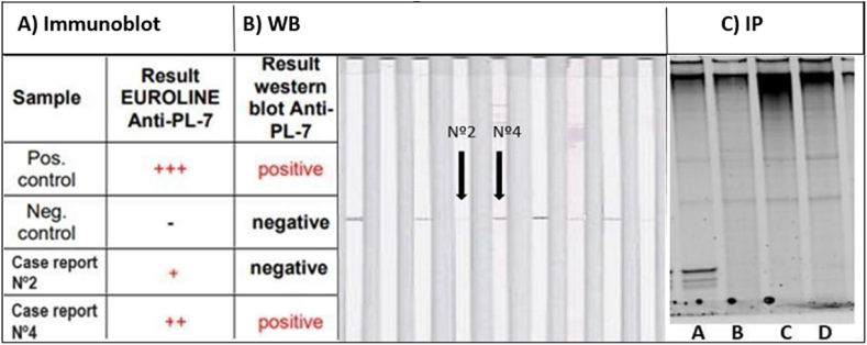
Cumulative evidence suggests a link between specific autoimmune diseases (AD), including idiopathic inflammatory myopathies (IIM), and SARS-CoV-2 infection or COVID-19 vaccination. Anti-synthetase syndrome (ASS), a subset of IIM, is defined by the presence of autoantibodies against aminoacyl-tRNA synthetase (anti-ARS) and is strongly associated with interstitial lung disease (ILD), a major contributor to severe complications and reduced survival. We present four clinical cases of patients who developed autoantibodies against threonyl (PL-7) and alanyl (PL-12) synthetases associated with ASS-ILD shortly after SARS-CoV-2 infection or COVID-19 vaccination. Anti-ARS autoantibodies were identified using three complementary methods: immunoblotting, western blotting (WB) and the method considered the gold standard, immunoprecipitation (IP), which ensures accurate interpretation of results. The study highlights the clinical and pathogenic overlap between ASS-ILD and SARS-CoV-2-related lung involvement.Both conditions share similar high-resolution computed tomography (HRCT) patterns, including inflammation and pulmonary fibrosis (PF), driven by IFN-γ signaling, which complicates accurate diagnosis. Our results provide novel insights into the temporal association of SARS-CoV-2 and vaccine exposure with ASS-ILD, focusing on possible molecular mimicry between viral proteins and ARS molecules as a potential mechanism. Understanding the involvement of specific anti-ARS autoantibodies (PL-7 and PL-12) and the identification of genetic predispositions (HLA-B∗08:01 and HLA-DRB1∗03:01) in these patients may be key to underpinning these autoimmune manifestations. The study underscores the importance of a multidisciplinary approach and vigilant follow-up to optimize diagnosis and management. Further research is essential to elucidate the causal relationships and molecular mechanisms behind these observations.
Keywords: Anti-synthetase syndrome; COVID-19; Idiopathic inflammatory myopathies; Interstitial lung disease; SARS-CoV-2 vaccine.
© 2024 Published by Elsevier Ltd.
Conflict of interest statement
The authors declare that they have no known competing financial interests or personal relationships that could have appeared to influence the work reported in this paper.
Figures


Similar articles
-
Analysis of the clinical features of antisynthetase syndrome: a retrospective cohort study in China.Clin Rheumatol. 2023 Mar;42(3):703-709. doi: 10.1007/s10067-022-06404-8. Epub 2022 Oct 29. Clin Rheumatol. 2023. PMID: 36308573
-
Increased Risk of Myositis-Specific and Myositis-Associated Autoantibodies After COVID-19 Pandemic and Vaccination: A Spanish Multicenter Collaborative Study.Biomedicines. 2024 Dec 10;12(12):2800. doi: 10.3390/biomedicines12122800. Biomedicines. 2024. PMID: 39767707 Free PMC article.
-
Anti-synthase syndrome associated with SARS-Cov-2 infection.BMC Pulm Med. 2024 Apr 15;24(1):179. doi: 10.1186/s12890-024-02966-2. BMC Pulm Med. 2024. PMID: 38622599 Free PMC article.
-
Antisynthetase syndrome and pulmonary hypertension: report of two cases and review of the literature.Mod Rheumatol Case Rep. 2021 Jan;5(1):152-155. doi: 10.1080/24725625.2020.1794521. Epub 2020 Jul 22. Mod Rheumatol Case Rep. 2021. PMID: 32697139 Review.
-
[Antisynthetase myopathy].Rinsho Shinkeigaku. 2020 Mar 31;60(3):175-180. doi: 10.5692/clinicalneurol.cn-001383. Epub 2020 Feb 26. Rinsho Shinkeigaku. 2020. PMID: 32101845 Review. Japanese.
References
-
- Cavagna L., Nuño L., Sciré C.A., et al. AENEAS collaborative group Serum Jo-1 autoantibody and isolated arthritis in the antisynthetase syndrome: a review of the literature and report of the experience of AENEAS collaborative group. Clin. Rev. Allergy Immunol. 2017;52:71–80. doi: 10.1007/s12016-016-8544-0. - DOI - PubMed
Publication types
LinkOut - more resources
Full Text Sources
Research Materials
Miscellaneous
