. 2025 Jan 21:13:1479960.
doi: 10.3389/fcell.2025.1479960. eCollection 2025.

Placental trophoblast aging in advanced maternal age is related to increased oxidative damage and decreased YAP

Affiliations

Placental trophoblast aging in advanced maternal age is related to increased oxidative damage and decreased YAP

Song Guo et al. Front Cell Dev Biol. .

Abstract

Introduction: The advanced maternal age (AMA) pregnancies escalate rapidly, which are frequently linked to higher risks of adverse outcomes. Advanced maternal age (AMA) placenta exhibited premature aging, presumably resulting in trophoblast dysfunction, inadequate placentation. However, the precise reasons and mechanisms of trophoblast aging in AMA placenta remain unclear, posing a significant limitation to provide effective guidance for prenatal healthcare in clinical settings. Notably, the organism shows heightened vulnerability to oxidative damage as it ages. YAP (Yes-associated protein) was reported to play a critical role in regulation of aging and resisting oxidative damage, yet these roles had not been elucidated in the placenta. Therefore, this study explored the relationship between trophoblast cell aging and oxidative injury and YAP in AMA pregnancy, which not only provided an insight into the mechanisms of trophoblast cell aging, but also provide valuable directions for healthcare during AMA pregnancy.

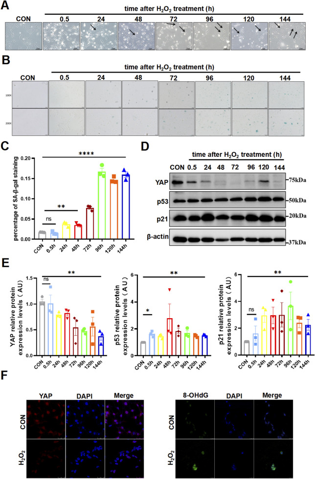
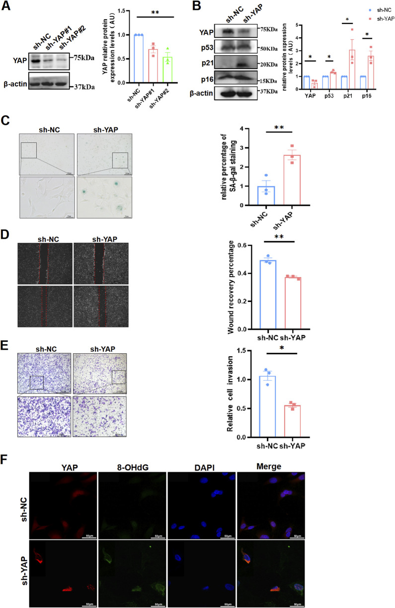
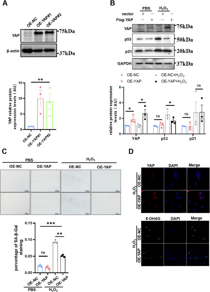
Methods: In this study, human term placentas were collected from AMA and normal pregnancies for the analysis of aging, oxidative damage and YAP level. HTR8/SVneo cells were manipulated with (hydrogen peroxide) H2O2 to explore the effects of oxidative damage on trophoblast cell senescence and YAP levels. YAP expression in HTR8/SVneo cells was manipulated to investigate its role in trophoblastic senescence and oxidative damage.

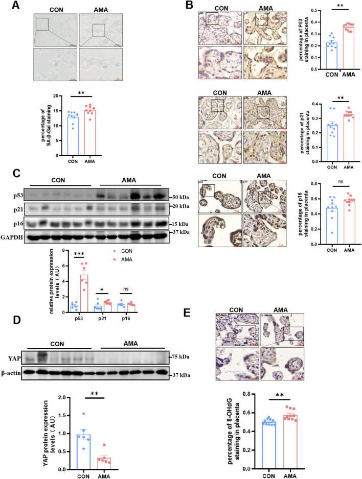
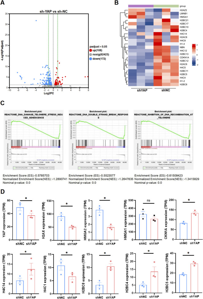
Results: Compared with the control group, the AMA placenta exhibits increased aging biomarkers, which is coupled with an elevation in oxidative damage within placental trophoblast cells and a notable decline in YAP levels. Cellular experiments demonstrated that oxidative damage from H2O2 triggered trophoblast cell senescence and resulted in a reduction of YAP levels. Furthermore, employing molecular modification to silence YAP expression in these cells led to an induction of aging. Conversely, overexpressing YAP ameliorated both trophoblast cell aging and the associated DNA oxidative damage that arised from H2O2.

Conclusion: The decline of YAP in AMA pregnancy should be responsible for the increased oxidative injury and premature placenta aging, indicating that YAP plays a significant role in combating oxidative damage and delaying aging, thereby providing a new guidance for prenatal care in AMA pregnancies. Maintaining YAP levels or implementing anti-oxidative stress interventions could potentially mitigate the incidence of complications involved AMA pregnancy.

Keywords: DNA oxidative damage; YAP; advanced maternal age; pregnancy complication; trophoblast aging.


Conflict of interest statement

The authors declare that the research was conducted in the absence of any commercial or financial relationships that could be construed as a potential conflict of interest.

Figures

FIGURE 1
FIGURE 1
AMA placentas showed a aging phenotype and were associated with decreased YAP and increased DNA oxidative damage. (A) SA-β-Gal staining of human term placenta sections (n = 10 patients per group, 3 random fields per patient). Scale bars, 100 μm. (B) IHC staining of p53, p21 and p16 in human term placentas (n = 10, 3 fields per patient). Scale bars, 50 μm. (C) Western blotting of p53, p21 and p16 protein expression in human term placentas (n = 6). (D) Western blotting of YAP protein expression in human term placentas (n = 6). (E) IHC staining of 8-OHdG in human term placentas (n = 10, 3 fields per patient). All data are presented as the mean ± SEM. *p < 0.05, **p < 0.01. Student’s t test. AU, arbitrary unit.
FIGURE 2
FIGURE 2
Oxidative stress induced senescence and attenuated YAP Expression in HTR8/SVneo cells. (A) HTR8/SVneo cells morphology changed over time by light microscopy after treated with H2O2 (n = 3). The arrow indicated the cells that have become larger and flattened in morphology, which was the characteristic of senescence. (B) SA-β-Gal staining in HTR8/SVneo cells by light microscopy over time after exposure of H2O2 (n = 3). Scale bars, 50 μm. (C) Statistical analysis diagram of the SA-β-Gal staining results in (B) (n = 3). (D) Western blotting of YAP, p53, and p21 protein expression in HTR8/SVneo cells over time after treated with H2O2 (n = 3). (E) Semi-quantitative statistical analysis diagram of various proteins of (D) (n = 3). (F) IF staining of YAP (red) and 8-OHdG (green) in different HTR8/SVneo cells; nuclei were counterstained with DAPI (blue). Scale bars, 100 μm (n = 3). All data are presented as the mean ± SEM. *p < 0.05, **p < 0.01, and ns, no significant. Student’s t-test for two groups, and one-way ANOVA for three or more groups. ns, nonsignificant; AU, arbitrary unit. All data are representative of three independent experiments.
FIGURE 3
FIGURE 3
YAP deficiency resulted in senescence and DNA oxidative damage in HTR8/SVneo cells, and compromised its invasiveness. (A) Western blotting of YAP expression in different HTR8/SVneo cells. sh-NC, negative control cells transfected with scramble shRNA; sh-YAP, cells transfected with shRNAs targeting YAP (n = 3). (B) Western blotting of YAP, p53, p21 and p16 expression in different HTR8/SVneo cells. Sh-YAP#1 was used in the following experiments (n = 3). (C) Representative SA-β-Gal staining in various HTR8/SVneo cells (n = 3). Scale bars, 100 μm. (D) Wound-healing assay of HTR8/SVneo cells (n = 3). Scale bars, 100 μm. (E) Transwell invasion assay of HTR8/SVneo cells (n = 3). Scale bars, 250 μm. (F) IF staining of YAP (red) and 8-OHdG (green) in HTR8/SVneo cells; nuclei were counterstained with DAPI (blue) (n = 3). Scale bar, 100 μm. All data are presented as the mean ± SEM. *p < 0.05, **p < 0.01, ***p < 0.001. Student's t test. AU, arbitrary unit. All data are representative of three independent experiments.
FIGURE 4
FIGURE 4
Supplement of YAP ameliorated aging and DNA oxidative damage induced by H2O2 in HTR8/SVneo cells. (A) Western blotting of YAP expression in different HTR8/SVneo cells (n = 3). OE-NC, negative control; OE-YAP, YAP overexpression. (B) Western blotting of YAP, p53, p21, and p16 expression in different HTR8/SVneo cells. OE-YAP#1 was used in the following experiments (n = 3). (C) Representative SA-β-Gal staining in various HTR8/SVneo cells (n = 3). Scale bars, 100 μm. OE-NC + H2O2, negative control cells treated with H2O2; OE-YAP + H2O2, YAP overexpression cells treated with H2O2. (D) IF staining of YAP (red) and 8-OHdG (green) in HTR8/SVneo cells; nuclei were counterstained with DAPI (blue) (n = 3). Scale bar, 100 μm. All data are presented as the mean ± SEM. *p < 0.05, **p < 0.01, ***p < 0.001, and ns, nonsignificant. Student’s t-test for two groups, and one-way ANOVA for three or more groups. AU, arbitrary unit. All data are representative of three independent experiments.
FIGURE 5
FIGURE 5
The transcriptome was altered in YAP-knockdown HTR8/SVneo cells. (A) Volcano plot of the significant differences in gene expression levels between sh-YAP and sh-NC HTR8/SVneo cells. sh-NC, negative control cells transfected with scramble shRNA; sh-YAP, cells transfected with shRNAs targeting YAP#1 (padj <0.05, n = 3). (B) Heat map of differentially expressed genes (DEGs) between sh-YAP and sh-NC by GESA analysis (padj <0.05, n = 3). (C) Multiple DNA damage related pathways were enriched including the DNA damage telomere stress induced senescence, DNA double-strand break pathways and inhibition of DNA recombination at telomere. (D) Further investigation of our RNA-seq data revealed that many genes involved in theses pathways were significantly changed in sh-YAP. *p < 0.05, and ns, nonsignificant. Student's t test.

Similar articles

References

    1. Beausejour C. M., Krtolica A., Galimi F., Narita M., Lowe S. W., Yaswen P., et al. (2003). Reversal of human cellular senescence: roles of the p53 and p16 pathways. EMBO J. 22, 4212–4222. 10.1093/emboj/cdg417 - DOI - PMC - PubMed
    1. Belo L., Caslake M., Santos-Silva A., Castro E. M., Pereira-Leite L., Quintanilha A., et al. (2004). LDL size, total antioxidant status and oxidised LDL in normal human pregnancy: a longitudinal study. Atherosclerosis 177, 391–399. 10.1016/j.atherosclerosis.2004.07.023 - DOI - PubMed
    1. Bruno R. M., Duranti E., Ippolito C., Segnani C., Bernardini N., Di Candio G., et al. (2017). Different impact of essential hypertension on structural and functional age-related vascular changes. Hypertension 69, 71–78. 10.1161/HYPERTENSIONAHA.116.08041 - DOI - PubMed
    1. Burton G. J., Jauniaux E. (2004). Placental oxidative stress: from miscarriage to preeclampsia. J. Soc. Gynecol. Investig. 11, 342–352. 10.1016/j.jsgi.2004.03.003 - DOI - PubMed
    1. Campisi J., d'Adda di Fagagna F. (2007). Cellular senescence: when bad things happen to good cells. Nat. Rev. Mol. Cell Biol. 8, 729–740. 10.1038/nrm2233 - DOI - PubMed

LinkOut - more resources