. 2025 Jan;12(Suppl 1):S14606.
doi: 10.1117/1.NPh.12.S1.S14606. Epub 2025 Feb 4.

Neurovascular coupling over cortical brain areas and resting state network connectivity with and without rigidified carotid artery

Affiliations

Neurovascular coupling over cortical brain areas and resting state network connectivity with and without rigidified carotid artery

Marleen E Bakker et al. Neurophotonics. 2025 Jan.

Abstract

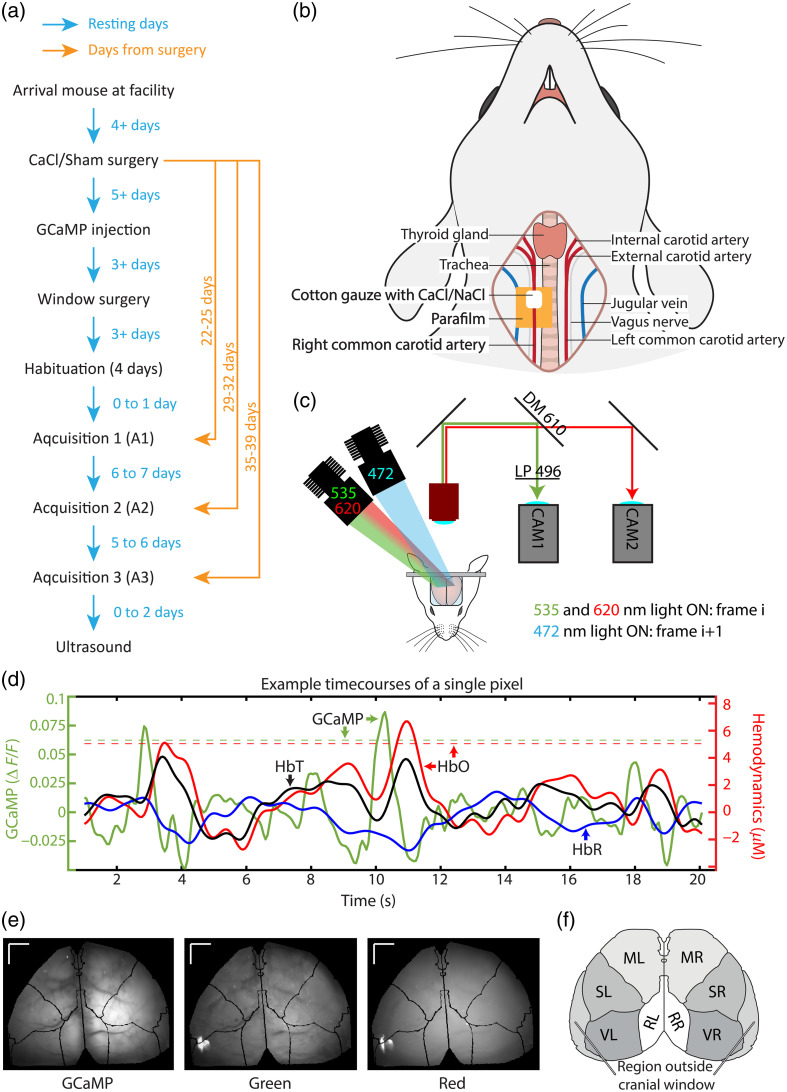
Significance: Neurovascular coupling (NVC) is key to research as hemodynamics can reflect neuronal activation and is often used in studies regarding the resting state network (RSN). However, several circumstances, including diseases that reduce blood vessel elasticity, can diminish NVC. In these cases, hemodynamic proxies might not accurately reflect the neuronal RSN.

Aim: We aim to investigate in resting state if (1) NVC differs over brain regions, (2) NVC remains intact with a mild rigidification of the carotid artery, (3) hemodynamic-based RSN reflects neuronal-based RSN, and (4) RSN differs with a mildly rigidified artery.

Approach: We rigidified the right common carotid artery of mice ( n = 15 ) by applying a CaCl 2 -soaked cloth to it (NaCl for Sham, n = 17 ). With simultaneous GCaMP and intrinsic optical imaging, we compared neuronal activation to hemodynamic changes over the entire cortex.

Results: NVC parameters did not differ between the CaCl and Sham groups. Likewise, GCaMP and hemodynamic RSN showed similar connections in both groups. However, the parameters of NVC differed over brain regions. Retrosplenial regions had a slower response and a higher HbR peak than sensory and visual regions, and the motor cortex showed less HbO influx than sensory and visual regions.

Conclusions: NVC in a resting state differs over brain regions but is not altered by mild rigidification of the carotid artery.

Keywords: GCaMP; intrinsic optical imaging; neurovascular coupling; resting state.


Figures

Fig. 1
Fig. 1
(a) Overview of the procedures that the mice were subjected to. (b) Schematic overview of the carotid artery calcification surgery. (c) Schematic overview of the imaging system. DM, dichroic mirror; LP, long pass filter. (d) Timecourses of a single pixel in the right sensory area of a single mouse. The GCaMP timecourse (in green) follows the y-axis on the left, whereas the hemodynamic timecourses (oxygenated hemoglobin—HbO—in red, deoxygenated hemoglobin—HbR—in blue, and total hemoglobin—HbT—in black) are on the right y-axis. The threshold for detecting spontaneous activation (z-score>1.95) for this pixel is depicted as a green dotted line for GCaMP, and a red dotted line for HbO. (e) Average images of the GCaMP, green, and red channels of a single acquisition. Black lines show the fitted Allen atlas. Lines in the top left corner depict the scale - lines are 1 mm. (f) Overview of the different regions of interest of the brain. VR, right visual area; SR, right sensory area; MR, right motor area; RR, right retrosplenial area; VL, left visual area; SL, left sensory area; ML, left motor area; RL, left retrosplenial area.
Fig. 2
Fig. 2
(a) Neurovascular coupling in individual mice. Each line depicts the average GCaMP, HbO, and HbR fluctuations 5 s before to 10 s after a detected spontaneous GCaMP activation. The green line depicts the GCaMP signal and corresponds to the left y-axis. Red lines depict HbO, and blue lines depict HbR, both corresponding to the right y-axis. Individual HbT curves are not shown to increase visibility. (b) Group average of the mice depicted in panel (a). The dark line shows the group average, and the lighter patches around the line show the standard error of the mean (SEM). See Figs. 10(b)–10(d) in Appendix E for plots with unfiltered data.
Fig. 3
Fig. 3
(a) Average curves for GCaMP (green), HbO (red), HbR (blue), and HbT (black) over different brain regions in the first acquisition of the Sham group. The left (green) y-axis depicts ΔF/F values for GCaMP, ranging from 0.05 to 0.05. The right (red) y-axis shows ΔμM for HbO, HbR, and HbT values, ranging from 2 to 2. The x-axis shows time, ranging from 5 to 5 s, with a notch at 0, where spontaneous GCaMP activation was detected. (b) Visual illustration of how the parameters in panel (c) were calculated. (c) Parameters related to NVC in different brain areas in the first acquisition of the Sham group. Letters above boxplots indicate significance: if two groups do not carry at least one similar letter, they differ significantly (p<0.05, see Table 1 in Appendix D).
Fig. 4
Fig. 4
Neurovascular coupling for all Sham mice of Acquisition 1 over the whole brain. The green line depicts the GCaMP signal and corresponds to the left y-axis. Red lines depict HbO, blue lines depict HbR, and black lines depict HbT, all corresponding to the right y-axis. The dark line shows the group average, and the lighter patches around the line show the standard error of the mean (SEM). (a) Curves of detected spontaneous GCaMP activations, where 0 is the time of detection based on GCaMP data. Note that these data are the same as shown in Fig. 2(b), with adjusted y-axes to make a direct comparison to panel (b). (b) Curves of detected spontaneous HbO activations, where 0 is the time of detection based on HbO data.
Fig. 5
Fig. 5
Parameters related to NVC in CaCl versus Sham-treated mice over three acquisitions. Values were taken from the right hemisphere. The same parameters as in Fig. 3 were calculated. None of the parameters showed a significant difference between the groups (Table 2 in Appendix G). For differences per brain area, see Fig. 11 in Appendix F.
Fig. 6
Fig. 6
(a) Average correlation scores between seeds in different brain areas of the CaCl group (top row) and Sham group (bottom row) for GCaMP (left), HbO (middle), and HbR (right) during acquisition 1 (other acquisitions can be found in Figs. 12 in Appendix H and Fig. 13 in Appendix I). (b) The differences between the average correlation scores of the Sham group minus the CaCl group over the three acquisitions (rows). None of the datatypes (GCaMP, HbO, and HbR) nor acquisition times showed a significant difference between the Sham and CaCl groups (Tables 3 and 4 in Appendix J).
Fig. 7
Fig. 7
Number of frames where the mouse was moving on the treadmill. Acquisitions existed of 9000 frames in total. Movement did not differ significantly between groups in the GCaMP acquisitions (A1: p=0.37, A2: p=0.39, A3: p=0.32), as tested with Kruskal–Wallis tests.
Fig. 8
Fig. 8
Number of spontaneous activations per brain region during acquisition 1, detected on GCaMP data (a) or HbO data (b). Per detected GCaMP activation, the percentage activations that were followed by the HbO timecourse reaching the detection threshold within 2 s are shown in panel (c). Conversely, panel (d) shows the percentage of detected HbO activations that were preceded (within 2 s) by a GCaMP peak that crossed the detection threshold. (e) The number of detected GCaMP activations of panel (a) was taken, but timepoints were randomized to fabricate a control group for panel (c). (f) Similar situation to panel (e), where the number of HbO-detected activations from panel (b) were randomized and checked for a corresponding GCaMP activation.
Fig. 9
Fig. 9
(a) Representative frame of an ultrasound recording in m-mode. (b) Diameter change of the right carotid artery. Dots indicate individual samples. N=7 for the CaCl group, and N=6 for the sham group. p=0.04.
Fig. 10
Fig. 10
GCaMP (depicted in green) corresponds to the left y-axis, whereas hemodynamics (HbO in red and HbR in blue) correspond to the right y-axis. The x-axis shows time in seconds, with 0 being the time of the detected spontaneous GCaMP activation. (a) Curves of GCaMP and hemodynamics per mouse, per region of interest. For clarity, HbT curves are not depicted. Each line shows the average curve over a region of interest of one mouse. (b) Curves of GCaMP and hemodynamics of one mouse, over the whole brain, with filtered and unfiltered data. (c) Neurovascular coupling curves per mouse. Unfiltered version of Fig. 2(a). (d) Group average of mice depicted in panel (c). The dark line shows the group average, and the patches show the standard error of the mean (SEM). Unfiltered version of Fig. 2(b).
Fig. 11
Fig. 11
Neurovascular coupling parameters for all brain areas over the three acquisitions. For statistics on the difference between CaCl and Sham groups over the right hemisphere, see Table 2 in Appendix G.
Fig. 12
Fig. 12
Correlation matrices of GCaMP (a), HbO (b), and HbR (c). Rows depict different acquisitions (A1, A2, and A3 for weeks 3, 4, and 5 after surgery, respectively). The first column shows the average correlation between seeds of the CaCl group, the second column shows that of the Sham group, and the third column shows the difference between Sham and CaCl (CaCl minus Sham average correlation). Differences were not significant (see Table 3 in Appendix J for statistics).
Fig. 13
Fig. 13
Correlation matrices of GCaMP (a), HbO (b), and HbR (c) with GSR. Rows depict different acquisitions (A1, A2, and A3 for weeks 3, 4, and 5 after surgery, respectively). The first column shows the average correlation between seeds of the CaCl group, the second column shows that of the Sham group, and the third column shows the difference between Sham and CaCl (CaCl minus Sham average correlation).

Similar articles

Cited by

References

    1. van den Heuvel M. P., Hulshoff Pol H.E., “Exploring the brain network: a review on resting-state fMRI functional connectivity,” Eur. Neuropsychopharmacol. 20(8), 519–534 (2010).EURNE810.1016/j.euroneuro.2010.03.008 - DOI - PubMed
    1. De Luca M., et al. , “fMRI resting state networks define distinct modes of long-distance interactions in the human brain,” Neuroimage 29(4), 1359–1367 (2006).NEIMEF10.1016/j.neuroimage.2005.08.035 - DOI - PubMed
    1. Biswal B., et al. , “Functional connectivity in the motor cortex of resting human brain using echo-planar MRI,” Magn. Reson. Med. 34(4), 537–541 (1995).MRMEEN10.1002/mrm.1910340409 - DOI - PubMed
    1. Fox M. D., Raichle M. E., “Spontaneous fluctuations in brain activity observed with functional magnetic resonance imaging,” Nat. Rev. Neurosci. 8(9), 700–711 (2007).NRNAAN10.1038/nrn2201 - DOI - PubMed
    1. Fox M. D., et al. , “The human brain is intrinsically organized into dynamic, anticorrelated functional networks,” Proc. Natl. Acad. Sci. U. S. A. 102(27), 9673–9678 (2005).10.1073/pnas.0504136102 - DOI - PMC - PubMed