. 2025 Mar 18;99(3):e0172424.
doi: 10.1128/jvi.01724-24. Epub 2025 Feb 5.

Usutu virus-induced meningoencephalitis in immunocompetent mice is characterized by the recruitment of mononuclear cells and a proinflammatory T helper 1 response

Affiliations

Usutu virus-induced meningoencephalitis in immunocompetent mice is characterized by the recruitment of mononuclear cells and a proinflammatory T helper 1 response

Rebeca Froes Rocha et al. J Virol. .

Abstract

Usutu virus (USUV) is an arbovirus and has emerged as a potential cause of encephalitis in humans and other vertebrates. The increasing detection of USUV in mosquitoes and birds across Africa and Central Europe, along with the lack of specific treatments or vaccines for many encephalitic orthoflaviviruses, underscores the need for focused research. In this study, we developed a USUV infection model in immunocompetent C57BL/6 mice (8-12 weeks old) to characterize disease development and associated inflammatory mechanisms. Mice were intracranially infected with 104 PFU of USUV, leading to neurological symptoms such as hunched posture, paralysis, conjunctivitis, and eventual death by day 6 post-infection. Meningeal cell infiltration and microglia activation were most prevalent in mouse brains; however, neuronal loss was not observed at the peak of the disease, which coincided with increased viral load and leukocyte infiltration. The immune response in the brain was marked by the systematic recruitment and activation of macrophages, neutrophils, and T lymphocytes. A noticeable shift was seen in CD4+ T cells toward T helper 1 (Th1) polarization, which corroborates a massive increase in the expression of Th1-associated cytokines and chemokines at the peak of infection, indicative of an augmented proinflammatory state. Additionally, a rise in regulatory T cells was observed, peaking on day 6 post-infection. These findings highlight the dynamic nature of the host response to USUV infection, enhance our understanding of the disease pathogenesis, and address the scarcity of immunocompetent experimental models for the investigation of neglected emerging flaviviruses.IMPORTANCEMosquito-borne viruses, including USUV, are maintained in nature through complex cycles involving arthropod vectors and vertebrate hosts. A comprehensive understanding of USUV biology and host-pathogen interactions is crucial for developing effective treatments, which necessitates reliable experimental models (G. J. Sips, J. Wilschut, and J. M. Smit, Rev Med Virol 22:69-87, 2012, https://doi.org/10.1002/rmv.712; T. C. Pierson and M. S. Diamond, Nat Microbiol 5:796-812, 2020, https://doi.org/10.1038/s41564-020-0714-0). The establishment of a USUV infection model in immunocompetent adult mice brings new perspectives on the inflammatory component of viral encephalitis, which is difficult to study in mice lacking antiviral interferon responses. Moreover, USUV is an emerging viral disease lacking therapeutic and preventive measures. The interplay of USUV pathogenesis and the host's immune response indicates that lymphocytes and monocytes participate in USUV infection in this model and could be explored in search of treatments targeting immunopathogenic processes triggered by infection.

Keywords: Th1 response; Usutu virus; flavivirus; immunopathogenesis; leukocyte recruitment; mouse model; viral encephalitis.


Conflict of interest statement

The authors declare no conflict of interest.

Figures

Fig 1
Fig 1
USUV replicates in the brain of immunocompetent mice leading to lymphopenia and meningeal cell infiltration. (A)C57BL6 8-week-old mice were inoculated in the brain with 20 µL of vehicle for the mock group (gray circle) or 101 PFU (pink circle), 102 PFU (green circle), 103 PFU (blue), and 104 PFU (red circle) of USUV for lethality curve analysis. (B)C57BL6 8-week-old mice infection with 104 of USUV leads to lymphopenia with a significant decrease of lymphocytes (gray bar) in serum at day 6 post-infection. Polymorphonuclear cells (red bar), monocytes (black bar). (C)Usutu virus intracranial inoculation (104 PFU) results in virus replication in brain tissue at day 3 (pink bar and circles) and day 6 post-infection (red bar and circles). ****P < 0,0001 relative to the NI control.
Fig 2
Fig 2
USUV infection in the brain of C57Bl6 mice results in meningeal cell infiltration. (A–E)NI group, (F–J)simulated, (K–O)USUV at day 3 post-infection (p.i.). Panoramic images of a hemisphere in the coronal section at the level of the hippocampus with a ×5 objective. Detail in ×20 objective of the regions of meninges, dentate gyrus (hippocampus), fibers of the tract (corpus callosum), and vessels at the intersection of the thalamus-hippocampus. (P–T)Cellular infiltrate in the meninges and perivascular regions in the brain from USUV-infected mice at 6 days post-infection. (U–X) Inactivated USUV does not result in brain damage after inoculation. Scale bars 100 µm.
Fig 3
Fig 3
Usutu virus (USUV) is detected in the mouse brain by immunofluorescence on day 6 p.i. and induces morphological alterations in microglia. Indirect immunofluorescence assays in coronal sections of the C57BL/6 mouse brain infected with USUV or vehicle. (A) Immunoreactivity of the 4G2 antibody, pan-flavivirus, reveals the detection of USUV in specific cells of the thalamus and amygdalar nucleus, exclusively in brains 6 days after infection. 4G2 labeling in thalamus cells is evident on day 6 p.i., absent in mice from the mock group, and on day 3 p.i. Images were acquired using a Leica SP8 confocal microscope with a ×40 objective plus ×1.5 digital zoom. (B)Immunodetection of microglia using the anti-Iba1 antibody in cells located in the hypothalamus. In brains infected for 6 days, morphological changes indicative of the microglia activation phenotype are observed. Images were captured with a Leica SP8 confocal microscope using a ×40 objective. Nuclei are labeled in blue with DAPI, while the anti-rabbit IgG secondary antibody is conjugated with Alexa 647 fluorophore in red. Scale bars represent 50 µm.
Fig 4
Fig 4
Immunohistochemical evaluation of neuronal loss in brain slides. (A)Neuronal loss was assessed by NeuN immunostaining. Images (×20) were taken from the basolateral amygdala. DAPI is marked in blue and NeuN is marked in red. Scale = 127.9 µm. (B and C)One-way analysis of variance of NeuN immunostaining mean and integrated density showed no difference between non-infected (NI) and infected groups. Neuronal loss was assessed by (D) TUNEL and (E) FJC staining. Images (×20) were taken from the basolateral amygdala. DAPI is marked in blue; TUNEL is marked in red (no staining observed); and FJC is marked in green (no staining observed). Scale = 511.6 µm. NI, n = 3; mock, n = 3; USUV D3, n = 4; and USUV D6, n = 5.
Fig 5
Fig 5
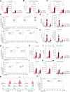
USUV induces leukocyte recruitment in brain tissue 6 days post-infection. Characterization of leukocyte populations, via flow cytometry, in brains of non-infected (NI) animals, mock-infected mice (inoculated with saline, gray bars) or infected with 104 PFU of USUV on day 3 (USUV D3) (pink bar) and day 6 (USUV D6) (red bar) post-infection. Leukocyte accumulation in the brain was characterized by (A)percentage of CD45+ live cells, (B)macrophages, (C)neutrophils. (D) Gating strategy for myeloid cell characterization and relative percentage of macrophage and neutrophil populations in CD45+ CD11b+ CD44+ cells. (E) Percentage of macrophage and neutrophil populations in CD45+ CD11b+ CD44+ cells live cells, (F) and expression of CXCR1 and CXCR2 in macrophages. (G)Gating strategy for lymphocytes and % relative of CD4+ and CD8+ in CD45+ CD3+ cells (H-I)Percentage and total cell count of T lymphocytes CD4+ and CD8+ in CD45+ CD3+ cells. (J-N)Expression of T-bet, CCR5, and CXCR3 in T CD4+ lymphocytes. (O)Expression of FoxP3 in T CD4+ CD25+ cells. The results are expressed as mean ± SD. *P < 0.05 relative to the NI/mock controls. Data are representative of one experiment and the following technical replicates: NI/mock (n = 4), USUV D3 (n = 3), and USUV D6 (n = 4).
Fig 6
Fig 6
USUV infection increases proinflammatory cytokines since day 3 p.i. in brain tissue. Detection of cytokine levels, via multiplex immunoassay, in brains of non-infected (NI) animals, mock-infected mice (inoculated with saline, gray bars) or infected with 104 PFU of USUV on day 3 (USUV D3) (pink bar) and day 6 (USUV D6) (red bar) p.i. Detected cytokine values were separated in graphs based on range of expression: (A)up to 80 pg/100 mg of tissue, (B)up to 600 pg/100 mg of tissue, (C)up to 12,000 pg/100 mg, or (D)up to 108 pg/100 mg. The results are expressed as mean ± SD. *P < 0.05, **P < 0.01, ***P < 0.001, ****P < 0.0001 relative to the NI/mock controls. Data are representative of two independent experiments and the following technical replicates: NI (n = 6), mock (n = 6), USUV D3 (n = 10), and USUV D6 (n = 9).

Similar articles

  • New Insights into the Susceptibility of Immunocompetent Mice to Usutu Virus.
    Benzarti E, Sarlet M, Franssen M, Desmecht D, Schmidt-Chanasit J, Garigliany MM. Benzarti E, et al. Viruses. 2020 Feb 8;12(2):189. doi: 10.3390/v12020189. Viruses. 2020. PMID: 32046265 Free PMC article.
  • Pathogenesis and shedding of Usutu virus in juvenile chickens.
    Kuchinsky SC, Frere F, Heitzman-Breen N, Golden J, Vázquez A, Honaker CF, Siegel PB, Ciupe SM, LeRoith T, Duggal NK. Kuchinsky SC, et al. Emerg Microbes Infect. 2021 Dec;10(1):725-738. doi: 10.1080/22221751.2021.1908850. Emerg Microbes Infect. 2021. PMID: 33769213 Free PMC article.
  • Differential neurovirulence of Usutu virus lineages in mice and neuronal cells.
    Clé M, Constant O, Barthelemy J, Desmetz C, Martin MF, Lapeyre L, Cadar D, Savini G, Teodori L, Monaco F, Schmidt-Chanasit J, Saiz JC, Gonzales G, Lecollinet S, Beck C, Gosselet F, Van de Perre P, Foulongne V, Salinas S, Simonin Y. Clé M, et al. J Neuroinflammation. 2021 Jan 6;18(1):11. doi: 10.1186/s12974-020-02060-4. J Neuroinflammation. 2021. PMID: 33407600 Free PMC article.
  • Usutu virus: an emerging flavivirus in Europe.
    Ashraf U, Ye J, Ruan X, Wan S, Zhu B, Cao S. Ashraf U, et al. Viruses. 2015 Jan 19;7(1):219-38. doi: 10.3390/v7010219. Viruses. 2015. PMID: 25606971 Free PMC article. Review.
  • Usutu virus: A new threat?
    Clé M, Beck C, Salinas S, Lecollinet S, Gutierrez S, Van de Perre P, Baldet T, Foulongne V, Simonin Y. Clé M, et al. Epidemiol Infect. 2019 Jan;147:e232. doi: 10.1017/S0950268819001213. Epidemiol Infect. 2019. PMID: 31364580 Free PMC article. Review.

Cited by

References

    1. Engel D, Jöst H, Wink M, Börstler J, Bosch S, Garigliany M-M, Jöst A, Czajka C, Lühken R, Ziegler U, Groschup MH, Pfeffer M, Becker N, Cadar D, Schmidt-Chanasit J. 2016. Reconstruction of the evolutionary history and dispersal of usutu virus, a neglected emerging arbovirus in europe and Africa. Mbio 7:e01938–15. doi:10.1128/mBio.01938-15 - DOI - PMC - PubMed
    1. Michel F, Sieg M, Fischer D, Keller M, Eiden M, Reuschel M, Schmidt V, Schwehn R, Rinder M, Urbaniak S, Müller K, Schmoock M, Lühken R, Wysocki P, Fast C, Lierz M, Korbel R, Vahlenkamp TW, Groschup MH, Ziegler U. 2019. Evidence for west nile virus and usutu virus infections in wild and resident birds in Germany, 2017 and 2018. Viruses 11:674. doi:10.3390/v11070674 - DOI - PMC - PubMed
    1. Lühken R, Jöst H, Cadar D, Thomas SM, Bosch S, Tannich E, Becker N, Ziegler U, Lachmann L, Schmidt-Chanasit J. 2017. Distribution of Usutu Virus in Germany and its effect on breeding bird populations. Emerg Infect Dis 23:1994–2001. doi:10.3201/eid2312.171257 - DOI - PMC - PubMed
    1. Clé M, Beck C, Salinas S, Lecollinet S, Gutierrez S, Van de Perre P, Baldet T, Foulongne V, Simonin Y. 2019. Usutu virus: a new threat? Epidemiol Infect 147:e232. doi:10.1017/S0950268819001213 - DOI - PMC - PubMed
    1. Ashraf U, Ye J, Ruan X, Wan S, Zhu B, Cao S. 2015. Usutu virus: an emerging flavivirus in Europe. Viruses 7:219–238. doi:10.3390/v7010219 - DOI - PMC - PubMed

MeSH terms

Supplementary concepts

LinkOut - more resources