. 2025 Mar 10;26(3):1748-1760.
doi: 10.1021/acs.biomac.4c01524. Epub 2025 Feb 5.

Simulation Study of the Water Ordering Effect of the β-(1,3)-Glucan Callose Biopolymer

Affiliations

Simulation Study of the Water Ordering Effect of the β-(1,3)-Glucan Callose Biopolymer

Robinson Cortes-Huerto et al. Biomacromolecules. .

Abstract

Callose, a polysaccharide closely related to cellulose, plays a crucial role in plant development and resistance to environmental stress. These functions are often attributed to the enhancement by callose of the mechanical properties of semiordered assemblies of cellulose nanofibers. A recent study, however, suggested that the enhancement of mechanical properties by callose might be due to its ability to order neighboring water molecules, resulting in the formation, up to room temperature, of solid-like water-callose domains. This hypothesis is tested by atomistic molecular dynamics simulations using ad hoc models consisting of callose and cellulose hydrogels. The simulation results, however, do not show significant crystallinity in the callose/water samples. Moreover, the computation of the Young's modulus gives nearly the same result in callose/water and in cellulose/water samples, leaving callose's ability to link cellulose nanofibers into networks as the most likely mechanism underlying the strengthening of the plant cell wall.


Conflict of interest statement

The authors declare no competing financial interest.

Figures
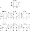
Figure 1
Figure 1
(a) Numbering of C atoms in the cyclic glucose molecule. The hydroxyl groups take the same number of the C they are bonded to. Comparison of the schematic structure of (b) callose and (c) cellulose polymers. Red line: glycosidic bonds. In (b) and (c), brackets identify the repeated unit in the biopolymers.
Figure 2
Figure 2
Snapshot of (a) the most dilute and (b) the most concentrated dispersion of callose in water. The two samples consist of the same number (9216) of glucose rings. (a) contains 51.9 water molecules per glucose ring and (b) contains 2.0 water molecules per glucose ring. Blue dots: O in water; red dots: O in callose; gray atoms: C in callose. Hydrogen atoms are not shown.
Figure 3
Figure 3
Close-contact between two callose chains in water, at relative concentration 85:15 callose/water wt %. To distinguish the two chains, all atom species were painted slightly differently on different chains.
Figure 4
Figure 4
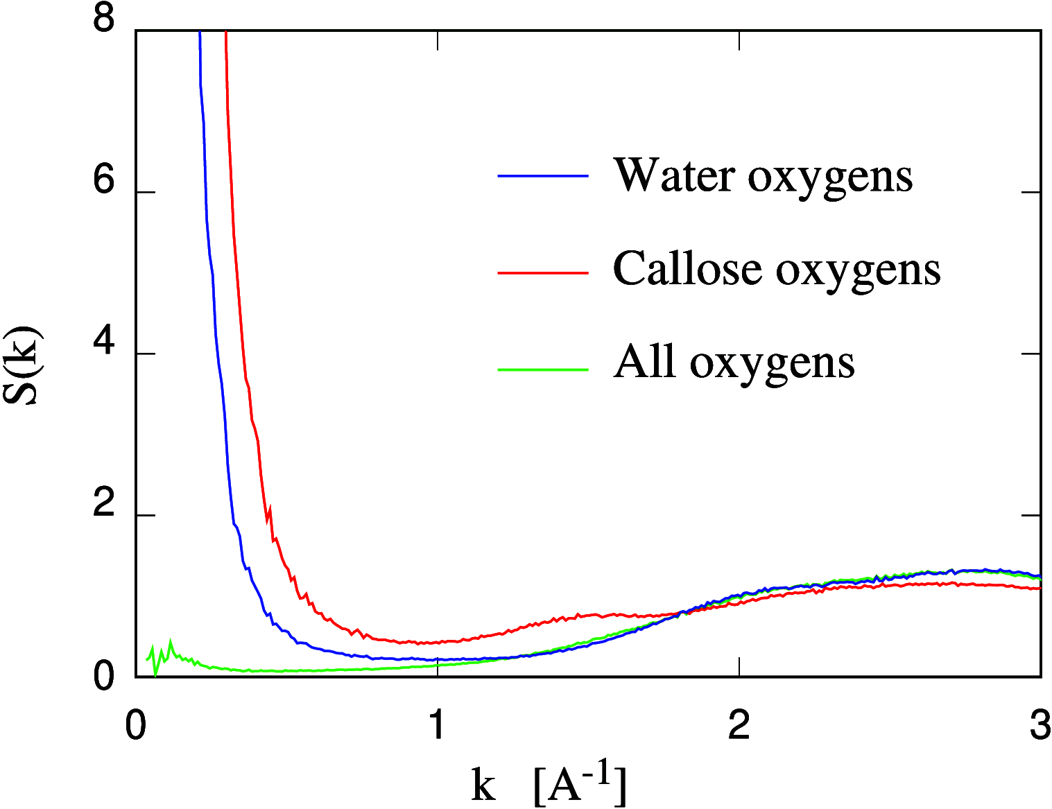
Structure factor computed for three different populations of oxygen atoms, as indicated in the labels and explained in the text. Data refer to the callose/water sample at 33.3 wt % callose concentration.
Figure 5
Figure 5
Water diffusion constant DW as a function of water concentration (in wt %) in the polysaccharide/water samples; The error bar on each DW value is comparable to the radius of the dots. The continuous lines are a guide to the eye. The red and blue dots close to the origin and not connected by the continuous lines represent two samples of very low water content (1 wt % water).
Figure 6
Figure 6
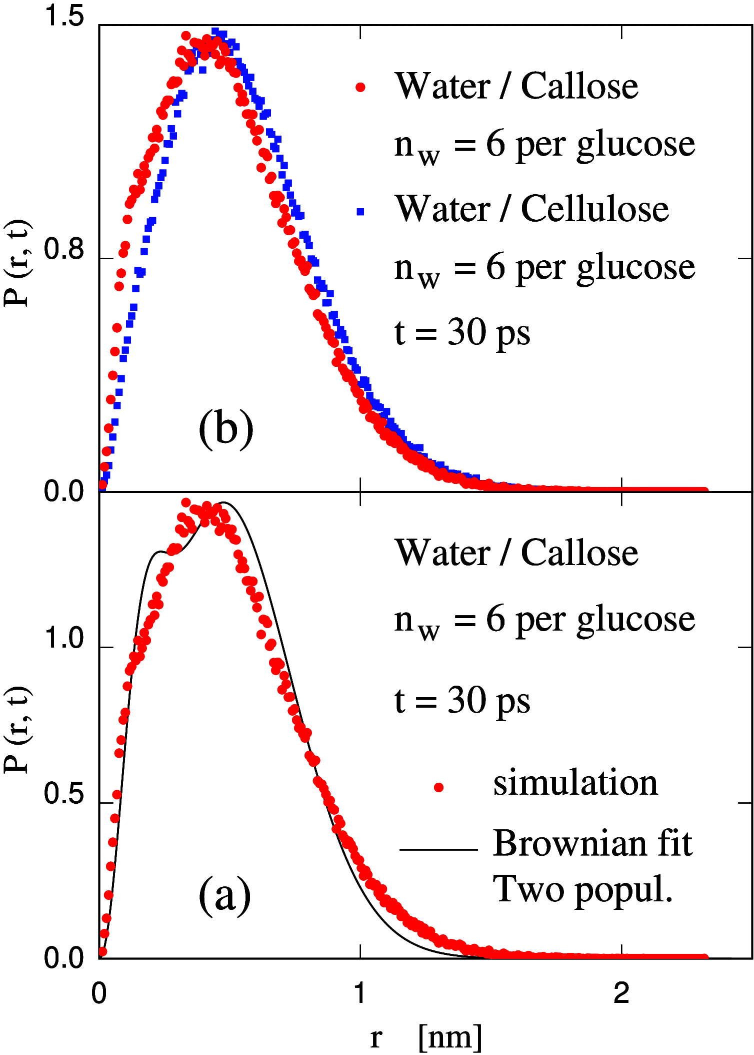
Probability distribution of water displacement over 30 ps. (a) Callose simulation data (dots) compared with the fit with two populations diffusing at different DW; (b) Comparison of the simulation data for callose/water (red dots) and cellulose/water (blue dots) samples.
Figure 7
Figure 7
Average number nHB of H-bonds per glucose unit for callose and cellulose chains in water as a function of the water wt % concentration in samples: red lines: callose/water; blue lines: cellulose/water; full lines: H-bonds donated by water to the polysaccharide chains; dash lines: H-bonds are donated by polysaccharides to water. Data represent averages over the last 400 ps of the production run for each system. The error bar is on the order of 2–3% of each value.
Figure 8
Figure 8
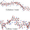
Snapshots of the most dilute (water concentration 85 wt %) callose/water and cellulose/water sample showing a segment of polysaccharide chain and the water molecules H-bonded to it. The two segments consist of nearly the same number of glucose units.
Figure 9
Figure 9
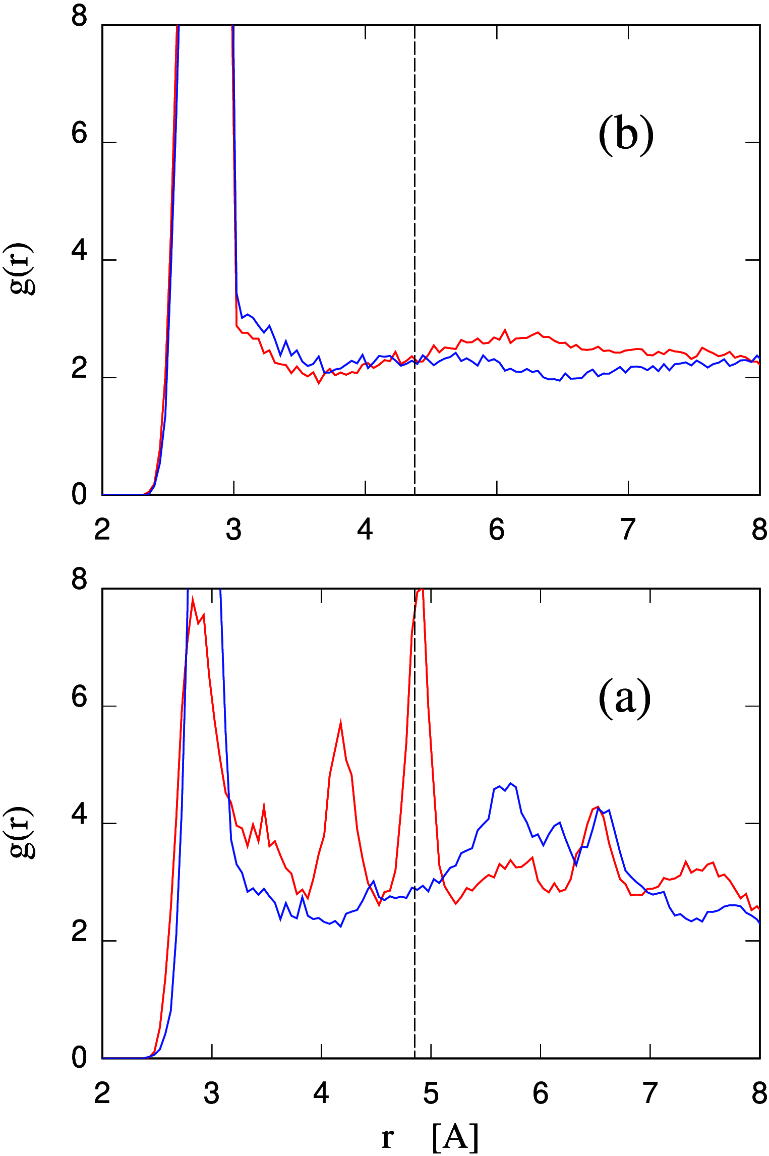
(a) Radial distribution function of hydroxyl oxygen atoms belonging to callose (red line) or cellulose (blue line) seen from a (average) hydroxyl oxygen atom of the same type at the origin. (b) Radial distribution function of oxygen atoms belonging to water molecules H-bonded to callose (red line) or cellulose (blue line) seen from an (average) hydroxyl oxygen atom belonging to the polysaccharide at the origin. All curves refer to the most dilute systems having a water concentration of 85 wt %. In both panels, the r = 4.85 Å radial distance is marked by a vertical dashed line (black).
Figure 10
Figure 10
Estimate of the Young’s modulus for the callose/water and cellulose/water samples of 60:40 polysaccharide/water wt % concentration. Dash lines: linear interpolation of the two sets of data. Estimate for water/callose: Y = 200 ± 30 MPa; cellulose/water: Y = 170 ± 15 MPa

Similar articles

References

    1. Galatis B.; Apostolakos P. A new callose function: involvement in differentiation and function of fern stomatal complexes. Plant Signal. Behav. 2010, 5, 1359–1364. 10.4161/psb.5.11.12959. - DOI - PMC - PubMed
    1. Hsieh Y. S.Y.; Kao M.-R.; Tucker M. R. The knowns and unknowns of callose biosynthesis in terrestrial plants. Carbohydr. Res. 2024, 538, 10910310.1016/j.carres.2024.109103. - DOI - PubMed
    1. Saito H.; Misaki A.; Harada T. A comparison of the structure of Curdlan and pachyman. Agr. Biol. Chem. 1968, 32, 1261–1269. 10.1080/00021369.1968.10859213. - DOI
    1. de Bary A. Recherches sur le développement de quelques champignons parasites. Annales des Sciences Naturelles, Botanique 1863, 20, 5–148.
    1. Ellinger D.; Voigt C. A. Callose biosynthesis in Arabidopsis with a focus on pathogen response: what we have learned within the last decade. Ann. Bot. 2014, 114, 1349–1358. 10.1093/aob/mcu120. - DOI - PMC - PubMed

LinkOut - more resources