. 2025 Feb 5;23(2):e3003001.
doi: 10.1371/journal.pbio.3003001. eCollection 2025 Feb.

Bacteriocin-like peptides encoded by a horizontally acquired island mediate Neisseria gonorrhoeae autolysis

Affiliations

Bacteriocin-like peptides encoded by a horizontally acquired island mediate Neisseria gonorrhoeae autolysis

Katy Poncin et al. PLoS Biol. .

Abstract

Neisseria gonorrhoeae is a human-specific pathogen that causes the important sexually transmitted infection, gonorrhoea, an inflammatory condition of the genitourinary tract. The bacterium is closely related to the meningococcus, a leading cause of bacterial meningitis. Both these invasive bacterial species undergo autolysis when in the stationary phase of growth. Autolysis is a form of programmed cell death (PCD) which is part of the life cycle of remarkably few bacteria and poses an evolutionary conundrum as altruistic death provides no obvious benefit for single-celled organisms. Here, we searched for genes present in these 2 invasive species but not in other members of the Neisseria genus. We identified a ~3.4 kb horizontally acquired region, we termed the nap island, which is largely restricted to the gonococcus and meningococcus. The nap island in the gonococcus encodes 3 cationic, bacteriocin-like peptides which have no detectable antimicrobial activity. Instead, the gonococcal Neisseria autolysis peptides (Naps) promote autolytic cell death when bacteria enter the stationary phase of growth. Furthermore, strains lacking the Naps exhibit reduced autolysis in assays of PCD. Expression of Naps is likely to be phase variable, explaining how PCD could have arisen in these important human pathogens. NapC also induces lysis of human cells, so the peptides are likely to have multiple roles during colonisation and disease. The acquisition of the nap island contributed to the emergence of PCD in the gonococcus and meningococcus and potentially to the appearance of invasive disease in Neisseria spp.


Conflict of interest statement

The authors have declared that no competing interests exist.

Figures

Fig 1
Fig 1. The nap island organisation and the absence of mNaps antimicrobial activity.
(A) Organisation of the nap island in Neisseria gonorrhoeae FA1090. Grey arrows, flanking genes (NEIS0808, pseudouridine synthase; NEIS0794, histidyl-tRNA synthase); blue arrows, genes coding for GG-containing peptides; orange arrows, genes predicted to be involved in peptide processing and secretion; green arrow, gene of unknown function; pink boxes, Correia elements. Percentage of GC content was plotted with Snap Gene Viewer. (B) Table detailing predicted function of genes in the nap locus. Details are available in S1 Fig. (C) Conservation of the nap island and flanking genes among Neisseria spp. Colours are scaled in percent of strain representatives possessing a homologue within each species. The data underlying this figure can be found in S2 Table. (D) mNaps (50 μm) were added to disks which were then placed on lawns of N. gonorrhoeae FA1090 (Ng), N. cinerea (Nc) CCUG 346T or 27178A, L. crispatus (Lc) ATCC 33820, and E. coli (Ec) MG1655 on solid media. Water and polymyxin B (50 μm) were used as negative and positive controls, respectively. Plates were incubated for 24 h and zones of growth inhibition (dotted lines) recorded. No growth inhibition was observed for any mNap (n = 3). Source data is in the file S1 Data. (E) MICs were determined by incubating bacteria in rich medium for 24 h with antibiotics (controls) or mNaps, starting from 256 μg/ml to 0.5 μg/ml in 2-fold dilutions. Bacteria were then spotted onto agar plates, which were incubated for 24 h. MBCs were the lowest concentration at which no growth was visible on plates. The mNaps did not inhibit the growth of any strain (shown by /, n = 3). The data underlying this figure can be found in S1 and S2 Data files.
Fig 2
Fig 2. Regulation of the nap island and the effect of mNaps on bacterial survival.
(A) RT-qPCR of genes in the nap island from N. gonorrhoeae FA1090 grown in GW liquid cultures. Samples were taken from flasks at different times (CFU, left panel) and mRNA levels of target genes determined by RT-qPCR (right panel). Gene expression results were normalised to samples at T0. (B) Bacteria were grown in 96-well plates in protein-free spermidine-free GW medium for 3 h before mNaps were added at various concentrations (arrow). At time points indicated, bacterial viability was determined by plating to solid media and incubation overnight. CFU were counted (dotted lines, limit of detection). Bottom panels represent the same data but grouped according to the growth phase (exponential and autolytic). Error bars, SD, two-way ANOVA (n = 4; p ≥ 0.033, not significant, not represented; p < 0.001, ***). The data underlying this figure can be found in S3 Data.
Fig 3
Fig 3. The nap island is involved in autolysis.
(A) Long-term survival of wild-type N. gonorrhoeae FA1090 and the ΔnapRABC mutant in the presence (arrow) or absence of mNaps (5 μm). The markerless ltgA deletion mutant was a control for a strain with reduced autolysis. Dotted lines, limit of detection. (B) Autolysis in rich medium. Bacteria were streaked on plates, grown overnight then resuspended in GCB at OD540 of ~0.3 and left in cuvettes. After 24 h at room temperature, bacteria were gently resuspended and the OD540 measured. One-way ANOVA with Dunnett’s multiple comparison against the wild-type strain (n = 3; p < 0.033, *; p < 0.002, **; p < 0.001, ***). The data underlying this figure can be found in S4 Data. (C) Autolysis in HEPES buffer without (left panel) or with (right panel) 10 mM MgCl2. A representative experiment is shown (last replicate is in the corresponding Suppl. source data files). (D) Transmission electron microscopy images from bacteria in HEPES (T = 75 min). Some wild-type bacteria appear normal (indicated with arrow numbered 1), with many others having a dense (arrow 2) or very dense (arrow 3) cytoplasm, periplasmic expansions (arrow 4), or appearing as ghost cells (arrow 5). Extracellular vesicles (arrow 6) and released cellular content (arrow 7) are also visible. Periplasmic expansions are not observable in the ΔltgA or ΔnapRABC strains. Cytoplasm condensation appears more homogenous in the ΔltgA mutant. The images underlying this figure can be found in https://doi.org/10.6084/m9.figshare.28077749.v1.
Fig 4
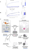
Fig 4. Effect of Naps on human red blood cells and proposed model.
(A) Different concentrations of mNaps were added to human RBCs for 1 h and the OD414 measured. Data were normalised to cytotoxicity with melittin (100%), or PBS (0%). Right panel, cytotoxicity induced by 1.56 μm of each mNap (n = 4; p < 0.002, **). (B) mNapA or mNapB were mixed with mNapC at a 1:1 ratio and cytotoxicity measured. Right panel, results with 3.15 μm of each peptide, n = 3, p < 0.033, *, one-way ANOVA with Dunnet’s multiple comparisons. The data underlying this figure can be found in S5 Data. (C) Proposed model of gonococcal autolysis. Peptidoglycan (PG) remodelling enzymes, including LtgA (pink) which localises at the septum, will release PG fragments that are recycled (purple arrows). When bacteria reach stationary phase, PG synthesis stops, but LtgA remains active, leading to the condensation of the cytoplasm (Fig 3D). Meanwhile, Naps are processed and secreted by NapP and NapF (orange), respectively, leading to their accumulation in the extracellular environment. Vacuolation is triggered, potentially through the action of inner membrane destabilising actors such as holins/toxins. Eventually, the outer membrane (OM) breaks when a critical concentration of mNaps (blue) is reached, leading to release of cellular contents and the appearance of ghost cells. For diplococci, cells undergoing PCD will benefit first-degree relatives by direct and indirect mechanisms.

Similar articles

References

    1. Quillin SJ, Seifert HS. Neisseria gonorrhoeae host adaptation and pathogenesis. Nat Rev Microbiol. 2018;16(4):226–240. doi: 10.1038/nrmicro.2017.169 - DOI - PMC - PubMed
    1. Wollstein M. Biological relationships of Diplococcus intrecallelularis and gonoccus. J Exp Med. 1907;9(5):588–605. - PMC - PubMed
    1. Flexner S. Contributions to the biology of Diplococcus intracellularis. J Exp Med. 1907;9(2):105–141. doi: 10.1084/jem.9.2.105 - DOI - PMC - PubMed
    1. Cloud KA, Dillard JP. A Lytic Transglycosylase of Neisseria gonorrhoeae Is Involved in Peptidoglycan-Derived Cytotoxin Production. Infect Immun. 2002;70(6):2752–2757. doi: 10.1128/IAI.70.6.2752-2757.2002 - DOI - PMC - PubMed
    1. Wegener WS, Hebeler BH, Morse SA. Cell envelope of Neisseria gonorrhoeae: relationship between autolysis in buffer and the hydrolysis of peptidoglycan. Infect Immun. oct 1977;18(1):210–219. doi: 10.1128/iai.18.1.210-219.1977 - DOI - PMC - PubMed

LinkOut - more resources