Arabidopsis KNL1 recruits type one protein phosphatase to kinetochores to silence the spindle assembly checkpoint
- PMID: 39908360
- PMCID: PMC11797493
- DOI: 10.1126/sciadv.adq4033
Arabidopsis KNL1 recruits type one protein phosphatase to kinetochores to silence the spindle assembly checkpoint
Abstract
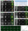
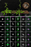
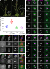
Proper chromosome segregation during cell division is essential for genomic integrity and organismal development. This process is monitored by the spindle assembly checkpoint (SAC), which delays anaphase onset until all chromosomes are properly attached to the mitotic spindle. The kinetochore protein KNL1 plays a critical role in recruiting SAC proteins. Here, we reveal that Arabidopsis KNL1 regulates SAC silencing through the direct recruitment of type one protein phosphatase (TOPP) to kinetochores. We show that KNL1 interacts with all nine TOPPs via a conserved RVSF motif in its N terminus, and this interaction is required for the proper localization of TOPPs to kinetochores during mitosis. Disrupting KNL1-TOPP interaction leads to persistent SAC activation, resulting in a severe metaphase arrest and defects in plant growth and development. Our findings highlight the evolutionary conservation of KNL1 in coordinating kinetochore-localized phosphatase to ensure timely SAC silencing and faithful chromosome segregation in Arabidopsis.
Figures


Similar articles
-
A coadapted KNL1 and spindle assembly checkpoint axis orchestrates precise mitosis in Arabidopsis.Proc Natl Acad Sci U S A. 2024 Jan 9;121(2):e2316583121. doi: 10.1073/pnas.2316583121. Epub 2024 Jan 3. Proc Natl Acad Sci U S A. 2024. PMID: 38170753 Free PMC article.
-
Three BUB1 and BUBR1/MAD3-related spindle assembly checkpoint proteins are required for accurate mitosis in Arabidopsis.New Phytol. 2015 Jan;205(1):202-15. doi: 10.1111/nph.13073. Epub 2014 Sep 29. New Phytol. 2015. Retraction in: New Phytol. 2016 Dec;212(4):1106. doi: 10.1111/nph.14225. PMID: 25262777 Retracted.
-
KNL1/Spc105 recruits PP1 to silence the spindle assembly checkpoint.Curr Biol. 2011 Jun 7;21(11):942-7. doi: 10.1016/j.cub.2011.04.011. Curr Biol. 2011. PMID: 21640906 Free PMC article.
-
How the SAC gets the axe: Integrating kinetochore microtubule attachments with spindle assembly checkpoint signaling.Bioarchitecture. 2015;5(1-2):1-12. doi: 10.1080/19490992.2015.1090669. Epub 2015 Oct 2. Bioarchitecture. 2015. PMID: 26430805 Free PMC article. Review.
-
Microtubule attachment and spindle assembly checkpoint signalling at the kinetochore.Nat Rev Mol Cell Biol. 2013 Jan;14(1):25-37. doi: 10.1038/nrm3494. Nat Rev Mol Cell Biol. 2013. PMID: 23258294 Free PMC article. Review.
Cited by
-
Functional characterization of the Csm1-like protein TITAN 9 in Arabidopsis thaliana.iScience. 2025 Jul 30;28(9):113251. doi: 10.1016/j.isci.2025.113251. eCollection 2025 Sep 19. iScience. 2025. PMID: 40837229 Free PMC article.
References
-
- Musacchio A., The molecular biology of spindle assembly checkpoint signaling dynamics. Curr. Biol. 25, R1002–R1018 (2015). - PubMed
-
- McAinsh A. D., Kops G. J. P. L., Principles and dynamics of spindle assembly checkpoint signalling. Nat. Rev. Mol. Cell Biol. 24, 543–559 (2023). - PubMed
-
- Hara M., Fukagawa T., Kinetochore assembly and disassembly during mitotic entry and exit. Curr. Opin. Cell Biol. 52, 73–81 (2018). - PubMed
-
- Ghongane P., Kapanidou M., Asghar A., Elowe S., Bolanos-Garcia V. M., The dynamic protein Knl1–A kinetochore rendezvous. J. Cell Sci. 127 (Pt. 16), 3415–3423 (2014). - PubMed
MeSH terms
Substances
LinkOut - more resources
Full Text Sources
Research Materials

