. 2025 Sep;145(9):2219-2228.e4.
doi: 10.1016/j.jid.2024.12.028. Epub 2025 Feb 3.

Pemphigus Vulgaris Autoantibodies Induce an Endoplasmic Reticulum Stress Response

Affiliations

Pemphigus Vulgaris Autoantibodies Induce an Endoplasmic Reticulum Stress Response

Coryn L Hoffman et al. J Invest Dermatol. 2025 Sep.

Abstract

Desmosomes are intercellular junctions that mediate cell-cell adhesion and are essential for maintaining tissue integrity. Pemphigus vulgaris (PV) is an autoimmune epidermal blistering disease caused by autoantibodies (IgG) targeting desmoglein 3, a desmosomal cadherin. PV autoantibodies cause desmosome disassembly and loss of cell-cell adhesion; however, the molecular signaling pathways that regulate these processes are not fully understood. Using high-resolution time-lapse imaging of live keratinocytes, we found that endoplasmic reticulum (ER) tubules make frequent and persistent contacts with internalizing desmoglein 3 puncta in keratinocytes treated with IgG of patients with PV. Biochemical experiments demonstrated that PV IgG activated ER stress signaling pathways, including both IRE1⍺ and PERK pathways, in cultured keratinocytes. Furthermore, ER stress transcripts were upregulated in the skin of patients with PV. Pharmacological inhibition of ER stress protects against PV IgG-induced desmosome disruption and loss of keratinocyte cell-cell adhesion, suggesting that ER stress may be an important pathomechanism and a therapeutically targetable pathway for PV treatment. These data support a model in which desmosome adhesion is integrated with ER function to serve as a cell adhesion stress sensor that is activated in blistering skin diseases.

Keywords: Cadherins; Desmosomes; ER stress; Endoplasmic reticulum; Pemphigus.


Conflict of interest statement

CONFLICT OF INTEREST

The authors state no conflict of interest.

Figures

Figure 1.
Figure 1.. ER tubules localize to desmosomes and remain associated with Dsg3 puncta during PV IgG-induced internalization.
N/TERT keratinocytes expressing luminal ER marker KDEL-StayGold (magenta) were pre-labeled with a fluorescently-conjugated (CF568) Dsg3 antibody (orange), treated with IgG, and imaged every 10 sec for various lengths of time during the first 3.5 hours following IgG addition. (a) Dsg3 puncta formed stable associations with peripheral ER tubules in NH IgG-treated cells even 3 hours after IgG addition (white arrows). (b) Stable Dsg3-ER associations were seen within the first 30 minutes following addition of PV1a IgG (white arrows). (c) ER tubules formed stable associations with Dsg3 puncta undergoing clustering (white arrows) and wrapped around linear arrays (yellow arrows) forming perpendicular to the cell-cell contact (dashed line). Scale bar = 5 μm.
Figure 2.
Figure 2.. ER stress is activated in PV IgG-treated keratinocytes and PV patient skin.
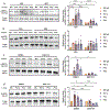
(a) NHEKs were treated with NH or PV IgG (PV1, PV2, or PV3) for 15 minutes to 1 hour, and ER stress marker phosphorylation was monitored by western blot. PV3 alone significantly increased IRE1α phosphorylation after 15 min. PV1 significantly increased eIF2α phosphorylation after 15 minutes and 1 hour, while PV2 significantly increased eIF2α phosphorylation after 1 hour. However, all PV IgG-treated NHEKs exhibited a trend towards increased IRE1α and eIF2α phosphorylation relative to NH IgG-treated NHEKs. (b) Expression of ER stress transcripts was monitored by qRT-PCR in PV patient skin biopsies from blister margins. All four patients show differential upregulation of ER stress markers relative to NH controls (dashed line). Mean value ± SEM is represented. ** P < 0.01, and *** P < 0.001 using an unpaired student’s t-test comparing PV IgG-treated to NH IgG-treated.
Figure 3.
Figure 3.. Miglustat and GSK2606414 prevent PV IgG-induced ER stress activation.
NHEKs were pre-treated with vehicle control (VEH), miglustat (MIG), DMSO, or GSK2606414 (GSK’414) and then treated with IgG from NH or PV patients (PV1, PV1a, PV2, or PV3) for 15 minutes or 1 hour, and ER stress marker phosphorylation was monitored by western blot. (a) At 15 minutes, MIG significantly prevented PV IgG-induced IRE1α phosphorylation but (b) did not have a significant effect on eIF2α phosphorylation. (c) GSK’414 significantly prevented PV IgG-induced phosphorylation of IRE1α at 15 minutes and (d) eIF2α at 1 hour. Mean value ± SEM is represented. * P < 0.05, ** P < 0.01, *** P < 0.001, and **** P < 0.0001 using a two-way ANOVA.
Figure 4.
Figure 4.. ER stress inhibitors prevent pathogenic PV IgG responses.
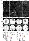
NHEKs were pre-treated with vehicle control (VEH), miglustat (MIG), DMSO, or GSK2606414 (GSK’414) and then treated with IgG from NH or PV patients (PV1a or PV2). (a) Localization of Dsg3 was monitored by immunofluorescence microscopy. Dsg3 border localization was decreased by both PV IgG in the VEH- and DMSO-treated cells but not in MIG- or GSK’414-treated cells. Scale bar = 20 μm. (b) Loss of adhesion was assessed by dispase fragmentation assay. PV IgG treatment significantly increased fragmentation relative to NH IgG-treated cells. MIG or GSK’414 treatment significantly reduced PV IgG-mediated fragmentation. Mean value ± SEM is represented. ns = not significant, * P < 0.05, *** P < 0.001, and **** P < 0.0001 using a two-way ANOVA.
Figure 5.
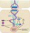
Figure 5.. Proposed model of ER stress signaling in response to PV IgG.
ER tubules (purple) form close associations with the desmosome junction (dark blue) and keratin filaments (light blue) on either side of a cell-cell contact. ER-desmosome associations are maintained following PV IgG-mediated Dsg3 clustering and linear array formation. Exposure to PV IgG leads to activation of ER stress through the IRE1α and PERK pathways, leading to transcriptional activation of the unfolded protein response and subsequent downstream signaling.

Update of

References

    1. Abeles I, Palma C, Meednu N, Payne AS, Looney RJ, Anolik JH. B Cell-Directed Therapy in Autoimmunity. Annu Rev Immunol 2024;42(1):103–26. - PubMed
    1. Abian O, Alfonso P, Velazquez-Campoy A, Giraldo P, Pocovi M, Sancho J. Therapeutic strategies for Gaucher disease: miglustat (NB-DNJ) as a pharmacological chaperone for glucocerebrosidase and the different thermostability of velaglucerase alfa and imiglucerase. Mol Pharm 2011;8(6):2390–7. - PubMed
    1. Alfonso P, Pampín S, Estrada J, Rodríguez-Rey JC, Giraldo P, Sancho J, et al. Miglustat (NB-DNJ) works as a chaperone for mutated acid β-glucosidase in cells transfected with several Gaucher disease mutations. Blood Cells, Molecules, and Diseases 2005;35(2):268–76. - PubMed
    1. Amagai M, Stanley JR. Desmoglein as a target in skin disease and beyond. J Invest Dermatol 2012;132(3 Pt 2):776–84. - PMC - PubMed
    1. Angulo-Urarte A, van der Wal T, Huveneers S. Cell-cell junctions as sensors and transducers of mechanical forces. Biochim Biophys Acta Biomembr 2020;1862(9):183316. - PubMed

MeSH terms

LinkOut - more resources