. 2025 Apr;134(4):1134-1145.
doi: 10.1016/j.bja.2024.11.045. Epub 2025 Feb 6.

Role of brain-derived neurotrophic factor in dysfunction of short-term to long-term memory transformation after surgery and anaesthesia in older mice

Affiliations

Role of brain-derived neurotrophic factor in dysfunction of short-term to long-term memory transformation after surgery and anaesthesia in older mice

Qiang Liu et al. Br J Anaesth. 2025 Apr.

Abstract

Background: Memory decline is one of the main manifestations in perioperative neurocognitive disorder. Short-term memory (STM) to long-term memory (LTM) transformation is one aspect of memory consolidation. Early-phase long-term potentiation (E-LTP) to late-phase long-term potentiation (L-LTP) is the molecular correlate of STM to LTM transformation. We examined whether the STM to LTM transformation was impaired after anaesthesia and surgery in older mice.

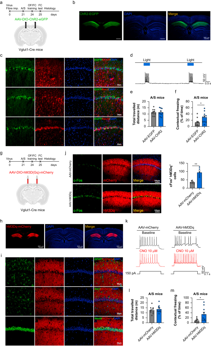
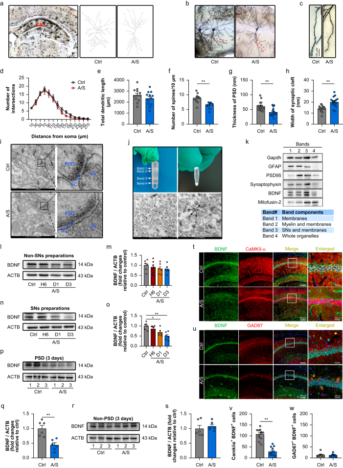
Methods: Optogenetics and chemogenetics were used to confirm the role of Vglut1+ glutamatergic neurones in the STM to LTM transformation in older mice. Synaptosomes were isolated to analyse expression of brain-derived neurotrophic factor (BDNF). Golgi-Cox staining and hippocampal field potential recordings were also used to measure synaptic plasticity.

Results: We found that the STM to LTM and E-LTP to L-LTP transformations were impaired after anaesthesia and surgery in older mice, and Vglut1+ excitatory neurone activity in the hippocampal CA1 region was reduced. BDNF expression decreased in the postsynaptic fraction, especially in Vglut1+ neurones, whereas cell-type specific overexpression of BDNF in Vglut1+ neurones reversed postoperative STM to LTM transformation dysfunction in older mice.

Conclusions: Reduced BDNF expression was involved in anaesthesia and surgery-induced impairment of the STM to LTM transition involving glutamatergic neurones in the hippocampal CA1 region of older mice. This provides a potential target that might be helpful for understanding and developing treatments for postoperative neurocognitive dysfunction.

Keywords: behavioural tagging; brain-derived neurotrophic factor; memory; perioperative neurocognitive dysfunction; synaptic tagging.


Conflict of interest statement

Declaration of interest The authors declare that they have no conflict of interest.

Figures

Fig 1
Fig 1
Anaesthesia and surgery-induced STM to LTM transformation in older mice. (a, b) Behaviour paradigm of STM to LTM transformation in normal older mice. n=10 per group. (c, d) Behaviour paradigm of STM to LTM transformation in normal older mice. n=10 per group. (e, f) STM was not impaired at days 1 and 3 after anaesthesia and surgery in older mice. (g, h) LTM transformation was impaired at days 1 and 3 after anaesthesia and surgery in older mice. n=10 per group. (i–l) Representative images (i) and quantification (j–l) of c-Fos-positive neurones in hippocampal CA1, CA3, and dentate gyrus (DG) subregions among the four groups. n=12 per group. (m and n) Schematic diagram and experimental scheme of E-LTP to L-LTP transformation. (o and p) E-LTP was not impaired after anaesthesia and surgery in CA1 of older mice. n=6 per group. (q, r) L-LTP was impaired after anaesthesia and surgery in CA1 of older mice. n=6 per group. A/S, anaesthesia and surgery; E-LTP, early-phase long-term potentiation; fEPSP, field excitatory postsynaptic potentials; L-LTP, late-phase long-term potentiation; LTM, long-term memory; OF, open field; STM, short-term memory. ∗ P<0.05, ∗∗ P<0.01.
Fig 2
Fig 2
Vglut1+ glutamatergic neurone activation was involved in the postoperative STM to LTM transformation dysfunction in older mice. (a, b) c-Fos+/Vglut1+ cell activation decreased in CA1 at day 3. n = 11–13 from four mice per group. (c, d) c-Fos+/Vgat+ GABAergic cell activation did not change significantly in CA1 at day 3. n=17–19 from five mice per group. (e) Whole-cell path recording picture. Action potential (AP) (f) firing rate (g), resting potential (h), and the threshold current (i) of Vglut1+ cells. n=12–14 neurones from three mice per group. A/S, anaesthesia and surgery; LTM, long-term memory; STM, short-term memory. ∗∗ P<0.01.
Fig 3
Fig 3
The role of Vglut1+ glutamatergic neurones in hippocampal CA1 during STM to LTM transformation for older mice. (a) Schema of the optogenetics approach flowchart. (b) eNpHR-mCherry expression in CA1. Scale bar (b): 500 μm; (c): 50 μm. (c) eNpHR mainly co-located with NeuN, not Iba1 or GFAP. (d) Yellow light inhibited action potentials in Vglut1+ neurones. Photoinactivation decreased contextual freezing time (f) without a change in total travelled distance (e). n=10 per group. (h) Schema of the chemogenetics approach flowchart. (j) CNO treatment inactivated fos+/Vglut1+ neurones after AAV-hM4D(Gi) injection in CA1 of Vglut1-Cre mice. Scale bar: 100 μm. (h) hM4D(Gi)-mCherry expression in CA1. (i) hM4D(Gi) mainly co-located with NeuN, not Iba1 or GFAP. (k) CNO perfusion inhibited action potential spiking. (l) The total travelled distance was not changed after CNO injection. (m) Contextual freezing time decreased after CNO injection. n=10 per group. Data are presented as mean (sem). A/S, anaesthesia and surgery; AP, action potential; CNO, clozapine N-oxide; FC, fear conditioning; LTM, long-term memory; OF, open field; STM, short-term memory. ∗P<0.05. ∗∗P<0.01.
Fig 4
Fig 4
Role of Vglut1+ glutamatergic neurones in hippocampus CA1 during postoperative STM to LTM transformation dysfunction in older mice. (a) Schema of the optogenetics approach flowchart. (b) ChR2-eGFP expression in CA1. Scale bar (b): 500 μm; (c): 50 μm. (c) ChR2 mainly co-located with NeuN, not Iba1 or GFAP. (d) Blue light activated action potentials in Vglut1+ neurones. Photoactivation prevented the decrease in contextual freezing time decreasing after anaesthesia and surgery in the AAV-ChR2 group compared with control group (f) without a change in total travelled distance (e). n=10 per group. (g) Schema of the chemogenetics approach flowchart. (j) CNO treatment activated fos+/Vglut1+ neurones after AAV-hM3D(Gq) injection in CA1 of Vglut1-Cre mice. Scale bar: 100 μm. (h) hM3D(Gq)-mCherry expression in CA1. (i) hM3D(Gq) mainly co-located with NeuN, not Iba1 or GFAP. (k) CNO perfusion activated action potential spiking. (l) Total travelled distance was not changed after CNO injection. (m) Contextual freezing time decrease after anaesthesia and surgery was attenuated after CNO injection in the AAV-h M3D(Gq) group. n=10 per group. Data are presented as mean (sem). A/S, anaesthesia and surgery; AP, action potential; CNO, clozapine N-oxide; FC, fear conditioning; LTM, long-term memory; OF, open field; STM, short-term memory. ∗P<0.05. ∗∗P<0.01.
Fig 5
Fig 5
Change in synaptic structural plasticity and the plasticity-related protein BDNF in synaptosomes after anaesthesia and surgery in older mice. (a–c) Synaptic structural plasticity changed in CA1 by Golgi staining. The number of spines was decreased in anaesthesia and surgery group (f) without a change in number of intersections (d) or total dendritic length (e). n=12 with three mice per group. Scale bar: 10 μm. (i) High magnification images by transmission electron microscopy show the synaptic structure changes. Scale bar: 500 nm. Analysis of the thickness of the postsynaptic density (PSD) (g) and width of the synaptic cleft (h). n=16–17 with three mice per group. (j) Synaptosome extraction and low power image by transmission electron microscopy showing the synaptosome. Scale bar: 2 μm or 500 nm. (k) Western immunoblot analyses of synaptosome preparations. (l) Western immunoblot showing BDNF levels in non-synaptosome preparations. (m) Analysis of BDNF in non- synaptosome preparations. n=6 per group. (n) Western immunoblot showing BDNF levels in synaptosome preparations. (o) Analysis of BDNF in synaptosome preparations. n=6 per group. (p) Western immunoblot showing BDNF levels in PSD fraction. (q) Analysis of BDNF in PSD fraction. n=6 per group. (r) Western immunoblot showing BDNF levels in non-PSD fraction. (s) Analysis of BDNF in non-PSD fraction. n=6 per group. Data are presented as mean (sem). (t, v) Image analysis showed that BDNF expression levels decreased in CaMKII+ excitatory neurones. (u, w) Image analysis showed that BDNF expression levels were not significantly changed in GAD67+ GABAergic neurones. A/S, anaesthesia and surgery; ACTB, beta-actin; BDNF, brain-derived neurotrophic factor; LTP, long-term plasticity; non-SNs, non-synaptosomes; SNs, synaptosomes. ∗P<0.05. ∗∗P<0.01.
Fig 6
Fig 6
Vglut1-specific overexpression of BDNF in CA1 improves STM to LTM transformation dysfunction of older mice after anaesthesia and surgery. (a) Diagram illustrating the injected virus and injection site in the CA1 region of the hippocampus. The inset shows expression of BDNF-eGFP in the CA1. Scale bar: 200 μm. (b, c) BDNF overexpression reversed the anaesthesia and surgery-induced decrease in BDNF mRNA and protein levels. n=6 per group. (d, e, g) Golgi–Cox staining, trait, and Sholl analyses among the four groups. (f–i) Golgi–Cox staining image and total dendritic length and number of spines analysed among the four groups. n=12 with three mice per group. (j) Transmission electron micrographs of four groups. (k, l) Analysis of PSD thickness and synaptic cleft width in the four groups. n=16–19 with three mice per group. (m, n) Tagging LTP in S2 and analysis of the fEPSP slope among the four groups. n=6 per group. (o) Contextual freezing time in the anaesthesia and surgery group was rescued by BDNF overexpression. n=10 per group. Data are presented as mean (sem). A/S, anaesthesia and surgery; ACTB, beta-actin; BDNF, brain-derived neurotrophic factor; fEPSP, field excitatory postsynaptic potentials; LTM, long-term memory; LTP, long-term plasticity; PSD, post-synaptic density; SC, synaptic cleft; STM, short-term memory; SV, synaptic vesicles.∗P<0.05. ∗∗P<0.01.

References

    1. Ackenbom M.F., Butters M.A., Davis E.M., Abebe K.Z., Baranski L., Zyczynski H.M. Incidence of postoperative cognitive dysfunction in older women undergoing pelvic organ prolapse surgery. Int Urogynecol J. 2021;32:433–442. - PMC - PubMed
    1. Mahanna-Gabrielli E., Zhang K., Sieber F.E., et al. Frailty is associated with postoperative delirium but not with postoperative cognitive decline in older noncardiac surgery patients. Anesth Analg. 2020;130:1516–1523. - PMC - PubMed
    1. Borchers F., Spies C.D., Feinkohl I., et al. Methodology of measuring postoperative cognitive dysfunction: a systematic review. Br J Anaesth. 2021;126:1119–1127. - PubMed
    1. Sungur Z., Şentürk M. Might complications of analgesic regimen affect cognitive assessment and how to diagnose POCD? Reg Anesth Pain Med. 2020;45:938–939. - PubMed
    1. Devinney M.J., Mathew J.P., Berger M. Postoperative delirium and postoperative cognitive dysfunction: two sides of the same coin? Anesthesiology. 2018;129:389–391. - PMC - PubMed

MeSH terms

Substances