. 2025 May;60(5):546-560.
doi: 10.1007/s00535-024-02210-y. Epub 2025 Feb 6.

Aldh2 and the tumor suppressor Trp53 play important roles in alcohol-induced squamous field cancerization

Affiliations

Aldh2 and the tumor suppressor Trp53 play important roles in alcohol-induced squamous field cancerization

Yuki Kondo et al. J Gastroenterol. 2025 May.

Abstract

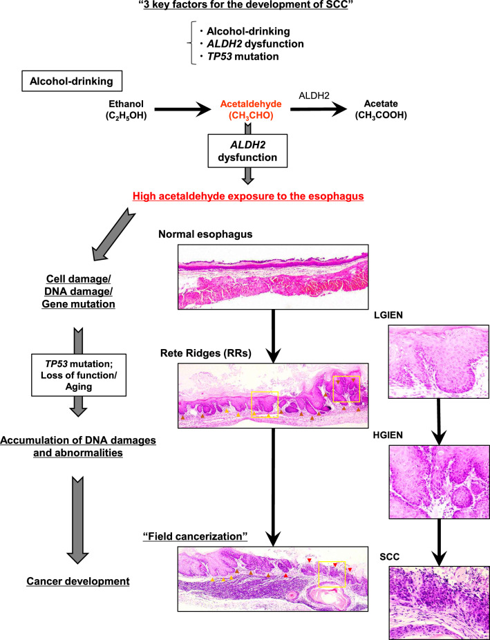
Background: Field cancerization defined by multiple development of squamous cell carcinomas (SCCs) in upper aerodigestive tract was explained by excessive alcohol intake. A dysfunctional mitochondrial aldehyde dehydrogenase 2 (Aldh2) delays the clearance of acetaldehyde, a genotoxic alcohol metabolite, and increases SCC risks. TP53 plays key roles in squamous carcinogenesis. However, the mechanism of alcohol-mediated squamous field cancerization has not been clearly elucidated.

Methods: We developed a novel genetically engineered mouse strain KTPA-/- (Krt5CreERT2; Trp53loxp/loxp; Aldh2-/-) featuring Aldh2-loss concurrent with epithelial-specific Trp53 deletion. These mice were given 10%-EtOH, and we evaluated the development of squamous cell carcinogenesis histologically and genetically.

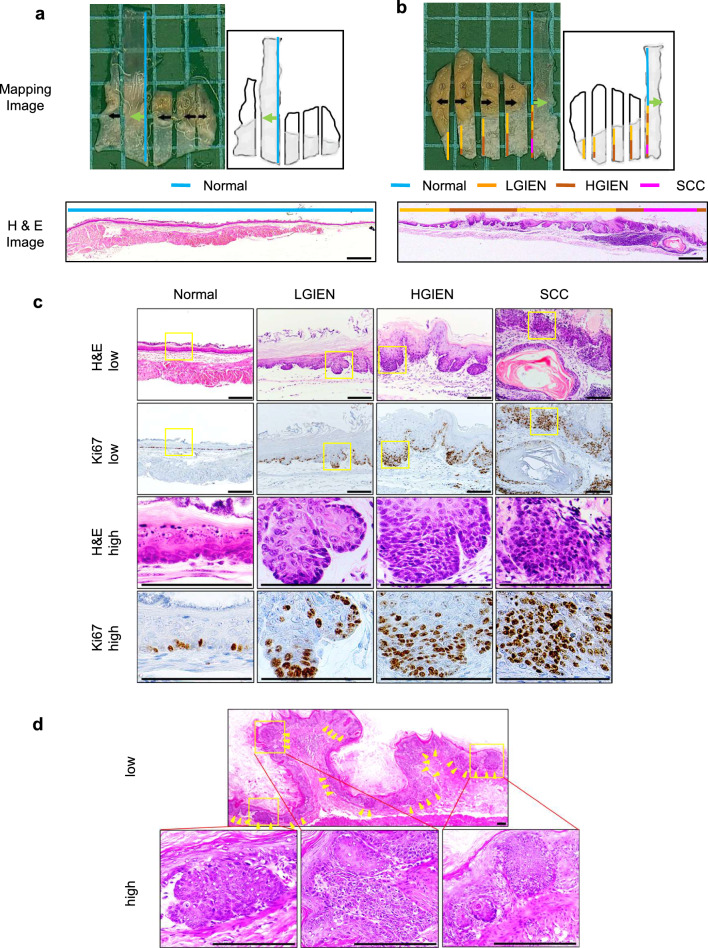
Results: Widespread multifocal rete ridges (RRs), characterized by downward growth of proliferative preneoplastic cells, were found only in Aldh2+/- and Aldh2-/- mice with keratin5-specific Trp53 deletion (KTPA+/- and KTPA-/- mice, respectively), and alcohol drinking apparently increased RR formation rate. SCC occurred only in KTPA-/- (Aldh2 loss/TP53 loss) mice with alcohol drinking (15/18: 83%). Total alcohol consumption volume was significantly higher in KTPA-/- (Aldh2 loss/TP53 loss) mice with SCCs than those without SCCs. Further, target sequence revealed the occurrence of genetic abnormalities including Trp53 mutations in the esophageal epithelium of Aldh2-/- mice with alcohol drinking, suggesting direct mutagenic effects of alcohol drinking to the esophageal epithelium.

Conclusion: This study provides for the first time the evidence that alcohol drinking, Aldh2 dysfunction and Trp53 loss cooperate in squamous field cancerization. Alcohol consumption volume affects the SCCs development, even in the same genotype.

Keywords: Alcohol-drinking; Aldh2; Field cancerization; SCC; TP53.


Conflict of interest statement

Declarations. Conflict of interest statement: All authors declare that they have no competing interests.

Figures

Fig. 1
Fig. 1
Alcohol drinking induces IEN and SCC in TAM-treated KTPA–/– (TP53–/–; Aldh2–/–) mice. a and b Representative macroscopic and microscopic images of dissected esophagus and forestomach samples were collected from TAM-treated KTPA–/– (TP53–/–; Aldh2–/–) mice that received drinking water containing no EtOH for 34 wk in a or 10% EtOH for 44 wk in (b). The specimens were grossed and serially sectioned in the direction indicated by black arrows. Each section was subjected to histopathological mapping of normal, low-grade IEN (LGIEN), high-grade IEN (HGIEN), and SCC lesions as indicated by a color code. Green arrows indicate directions of cross sections for the representative H&E-stained slides shown. c Representative H&E- and Ki67-stained slides of normal, LGIEN, HGIEN, and SCC lesions. The area demarcated by yellow rectangles was enlarged in the lower panels. d Multifocal IEN and invasive SCC lesions (yellow arrowheads). Invasive tumor fronts demarcated by yellow rectangles in the upper panel were enlarged in the lower panels. Scale bars = 0.5 mm in (a and b), and 100 µm in (c and d)
Fig. 2
Fig. 2
Rete ridges may be linked to mucosal inflammation during alcohol-induced carcinogenesis. a Representative H&E and immunohistochemistry images capturing rete ridges (RRs) (yellow arrowheads) characterized by highly proliferative Ki67 + cells and drop-shaped downward growth in the forestomach from alcohol-fed TAM-treated KTPA–/– (TP53–/–; Aldh2–/–) mice. Scale bars = 1 mm. b The number of RRs per 2 mm of the squamous epithelial basal segment is shown in the violin plots for all mouse strains and indicated treatment conditions, with the frequency for each group indicated at the top. c Inflammation scores plotted for all mouse strains and indicated treatment conditions. d Scatter plot showing the relationship between the number of RRs and inflammation scores of all strains and conditions. Red circles indicate the mice that developed IEN /SCC, whereas black circles indicate the mice with normal epithelium. n = 104, r = 0.6631, P < 0.0001, red circles vs black circles. The dotted lines represent the 95% confidence intervals for the linear regression
Fig. 3
Fig. 3
Alcohol consumption and esophageal carcinogenesis in KTPA–/– (TP53–/–; Aldh2–/–) mice. a Alcohol consumption during the initial 4 wk after the start of alcohol drinking was plotted for TAM-treated KTPA–/– (TP53–/–; Aldh2–/–) (n = 10), KTPA+/– (TP53–/–; Aldh2+/–) (n = 10), and KTPA+/+ (TP53–/–; Aldh2+/+) (n = 5) mice with indicated Aldh2 status. One-way ANOVA (P < 0.05), followed by post hoc Tukey’s multiple comparison test. Error bars represent mean ± SD. ****P < 0.0001. b The total alcohol amount consumed by TAM-treated KTPA–/– (TP53–/–; Aldh2–/–) (n = 10) was plotted for the entire alcohol-drinking period. 2-Tailed Student t test. Error bars represent mean ± SD. *P < 0.05. In (ab), each circle represents a single mouse. Red circles indicate mice with IEN or SCC mice, and black circles indicate mice with normal mucosa mice
Fig. 4
Fig. 4
TAM-treated and alcohol-fed KTPA–/– (TP53–/–; Aldh2–/–) mice induce SCC development at high rates by Cohort 2 study. a, b Representative H&E- and Ki67-stained images with SCC. The right panel were enlarged in the left panels. c, d Representative H&E- and Ki67-stained images of tumor area with strong inflammatory changes. The right panel were enlarged in the left panels. e, f Representative H&E- and Ki67-stained images of rete ridges (yellow arrowheads) with strong inflammatory changes. Scale bars = 100 µm (a, b, c, d, e and f). g Scatter plot showing the relationship between the number of RRs and inflammation scores of alcohol-fed TAM-treated KTPA–/– (TP53–/–; Aldh2–/–) mice. Red circles indicate the mice developed IEN/SCC, whereas black circles indicate the mice with normal epithelium. n = 18, r = 0.8160, P < 0.0001, red circles vs black circles
Fig. 5
Fig. 5
Effects of long-term acetaldehyde exposure on esophageal epithelial cells in vitro and effects of long-term alcohol drinking on Aldh2-deficient mice in vivo. EPC3-hTERT cells were cultivated for 24 weeks in a medium supplemented with or without 0.2 mM acetaldehyde (AA), and then those cells were subjected to colony formation assays. The average size (a) and number (b) of colonies were determined under the high-power field. n = 9, Error bars represent mean ± SD. **P < 0.01. Target sequencing of esophageal epithelium in Aldh2ko/ko mice treated with 10% EtOH or water (Control) for 3 months was conducted (Control, n = 3; EtOH, n = 3). C Total count of short variants (variant type-specific: single nucleotide variant (SNV), insertion (INS), deletion (DEL)) in 3 mice for 6 genes (Trp53, Notch1, Notch3, Pik3ca, Cdkn2a, and Fat1). d Total count of SNVs (base substitution pattern-specific) in 3 mice for 6 genes. e Total count of short variants in 3 mice for Trp53 coding regions. f Total count of SNVs in 3 mice for Trp53 coding regions. g Detection of three SNVs in the Trp53 coding regions in Aldh2-deficient mice with alcohol drinking for 3 months
Fig. 6
Fig. 6
Image of esophageal field carcinogenesis caused by alcohol. Pathophysiology of alcohol-induced SCC development. When alcohol is consumed, EtOH is metabolized to acetaldehyde, and the acetaldehyde is detoxified to acetic acid by aldehyde dehydrogenase 2 (ALDH2). If the ALDH2 gene polymorphism is impaired in this process, acetaldehyde is not degraded, resulting in exposure of the squamous epithelium to high concentrations of acetaldehyde. The mutagenicity of acetaldehyde causes cell damage, DNA damage, and gene mutations in the esophageal epithelium. The Trp53 mutation, a typical gene mutation, causes characteristic morphologic changes (Rete ridges: red arrowheads are SCC, brown arrowheads are HGIEN, orange arrowheads are LGIEN) in the esophageal epithelium as the gene mutation accumulates with age, and changes such as LGIEN and HGIEN occur. These lesions are multiple and eventually lead to invasive carcinoma. The squamous epithelium by yellow rectangles in the center panel were enlarged in the right panels. This condition may reflect field carcinization, indicating that alcohol consumption, Aldh2 deficiency, and Trp53 mutation are essential key factors in squamous field cancerization

References

    1. Slaughter DP, Southwick HW, Smejkal W. Field cancerization in oral stratified squamous epithelium; clinical implications of multicentric origin. Cancer. 1953;6:963–8. - PubMed
    1. Murai K, Dentro S, Ong SH, et al. p53 mutation in normal esophagus promotes multiple stages of carcinogenesis but is constrained by clonal competition. Nat Commun. 2022;13:6206. - PMC - PubMed
    1. Muto M, Hitomi Y, Ohtsu A, et al. Association of aldehyde dehydrogenase 2 gene polymorphism with multiple oesophageal dysplasia in head and neck cancer patients. Gut. 2000;47:256–61. - PMC - PubMed
    1. Muto M, Hironaka S, Nakane M, et al. Association of multiple Lugol-voiding lesions with synchronous and metachronous esophageal squamous cell carcinoma in patients with head and neck cancer. Gastrointest Endosc. 2002;56:517–21. - PubMed
    1. Baan R, Straif K, Grosse Y, et al. Carcinogenicity of alcoholic beverages. Lancet Oncol. 2007;8:292–3. - PubMed

MeSH terms