. 2025 Feb 5;16(1):1142.
doi: 10.1038/s41467-025-56217-x.

Testosterone exacerbates neutrophilia and cardiac injury in myocardial infarction via actions in bone marrow

Affiliations

Testosterone exacerbates neutrophilia and cardiac injury in myocardial infarction via actions in bone marrow

Elin Svedlund Eriksson et al. Nat Commun. .

Abstract

Men develop larger infarct sizes than women after a myocardial infarction (MI), but the mechanism underlying this sex difference is unknown. Here, we demonstrated that blood neutrophil counts post-MI were higher in male than female mice. Castration-induced testosterone deficiency reduced blood neutrophil counts to the level in females and increased survival post-MI. These effects were mimicked by Osterix-directed ablation of the androgen receptor in bone marrow (BM). Mechanistically, androgens downregulated the leukocyte retention factor CXCL12 in BM stromal cells. Post-hoc analysis of clinical trial data showed that neutrophilia was greater in men than women after reperfusion of first-time ST-elevation MI, and tocilizumab, an interleukin-6 receptor inhibitor, reduced blood neutrophil counts and infarct size to a greater extent in men than women. Our work reveals a previously unknown mechanism connecting testosterone with neutrophilia and MI injury via BM and identifies the importance of considering sex when developing anti-inflammatory strategies to treat MI.


Conflict of interest statement

Competing interests: K.B. has received lecture fees from Amgen, AstraZeneca, Boehringer, Novartis, NovoNordisk, Pharmacosmos, Pfizer, and consultant fees from AstraZeneca, Boehringer, Pharmacosmos, and Pfizer. L.G. has received lecture fees from AstraZeneca, Boehringer Ingelheim, Novartis, and Amgen. The remaining authors declare no competing interests.

Figures

Fig. 1
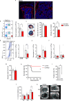
Fig. 1. Castration of male mice reduces neutrophilia, cardiac injury, and cardiac remodeling and increases survival after an acute MI.
ad Neutrophil counts in blood, area at risk (AAR) and MI size after 45 min ischemia followed by 24 h reperfusion. a, b Male and female mice; a *P = 0.038 (two-sided unpaired Student t test), n = 5 + 4; b ***P < 0.0001 (two-sided unpaired Student t test), n = 10 + 7. c, d Sham-castrated (control) and castrated (orchiectomized; ORX) male mice; c P = 0.057, n = 4 + 4 (two-sided Mann–Whitney); d **P = 0.0095 (two-sided Mann−Whitney), n = 4 + 6. eg MI was induced by permanent ligation of the left coronary artery in ORX or sham-castrated (control) mice. 48 h after MI or sham-MI (S-MI) surgery, neutrophils (e), monocytes (f), and macrophages (g) were quantified in the left ventricle (LV) by flow cytometry. e *P = 0.017 and **P = 0.004 (two-sided unpaired Student t test). eg n = 5 + 5 and 9 + 9 for S-MI and MI, respectively. hl MI was induced by permanent ligation of the left coronary artery in ORX or sham-castrated (control) mice. Echocardiography was performed before MI (baseline; BL) and 24 h and 3 weeks post-MI. h The area at risk analyzed at 24 h post-MI. i Survival rate; P = 0.050 (log-rank test). j Cardiac rupture rate during the 3 weeks of follow-up; *P = 0.044 (two-sided Fisher´s test); hj n = 18 + 14. LV volume in diastole at BL, 24 h and 3 weeks post-MI; n = 9 + 9 (BL), 10 + 12 (24 h), and 9 + 12 (3w), 3w, *P = 0.042 (two-sided unpaired Welch´s t test). l Representative echocardiographic images in sham-operated (control) and castrated (ORX) mice at 24 h and 3 weeks post-MI, scale bar = 1 mm. m, n Male mice were castrated and treated with vehicle (V) or testosterone (T) for 3 weeks before permanent ligation of the left coronary artery. 24 h after MI, plasma was assessed for myeloperoxidase (MPO), m *P = 0.050 (two-sided unpaired Student t test) and cardiac troponin I (cTnI), n; *P = 0.015 (two-sided unpaired Student t test); m, n; n = 8 + 9. Bars indicate means (a, b, eg, k, m, n) or medians (c, d), error bars are SEM (a, b, eg, k, m, n) or interquartile ranges (c, d), and circles represent individual mice. Source data are provided as Source data file.
Fig. 2
Fig. 2. Osterix-directed knockout of the androgen receptor reduces neutrophilia and cardiac remodeling and increases survival after an acute MI.
a A representative image of Osterix-Cre-directed tdTomato reporter signal in femur. Scale bars = 250 and 50 µm (zoomed image), respectively. b, c Neutrophil counts in blood, area at risk (AAR), and MI size after 45 min ischemia followed by 24 h reperfusion in O-ARKO and control (Osx1-Cre+) mice. b *P = 0.012 (two-sided unpaired Student t test); n = 6 + 5. c **P = 0.0061 (two-sided Mann–Whitney); n = 7 + 4. df MI was induced by permanent ligation of the left coronary artery in control and O-ARKO mice. Mice without MI surgery were used for reference. 48 h after the MI surgery, neutrophils (d), monocytes (e), and macrophages (f) were quantified in the left ventricle (LV) by flow cytometry. d **P = 0.0013 (two-sided unpaired Student t test), df n = 6 + 8 and 10 + 14 for no MI and MI, respectively. gk MI was induced by permanent ligation of the left coronary artery in O-ARKO or control mice. Echocardiography was performed before MI surgery (baseline; BL) and 24 h and 3 weeks post-MI. g The area at risk analyzed at 24 h post-MI; n = 11 + 12. h Survival rate; *P = 0.014 (log-rank test). i Cardiac rupture rate during the 3 weeks of follow-up; *P = 0.015 (two-sided Fisher´s test); hj n = 12 + 13. j LV volume in diastole at BL, 24 h and 3 weeks post-MI; n = 12 + 10 (BL), 11 + 12 (24 h), 6 + 11 (3w), 3w, *P = 0.048 (two-sided Mann–Whitney). k Representative echocardiographic images in sham-operated (control) and castrated (ORX) mice at 24 h and 3 weeks post-MI. Scale bar = 1 mm. Bars indicate means (b, dg, j) or medians (c), error bars are SEM (b, dg, j) or interquartile ranges (c) and circles represent individual mice. Source data are provided as Source data file.
Fig. 3
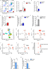
Fig. 3. Cytokine-controlled neutrophilia depends on the AR in BM stroma.
a, b Mice were injected i.p. twice daily with 125 µg/kg of G-CSF or vehicle on three consecutive days. Neutrophil (Ly6G+) counts in blood were measured by flow cytometry; representative flow plots in a. a Vehicle or G-CSF injection in control or O-ARKO mice; *P = 0.034 (two-sided unpaired Student t test); n = 3 + 5 and 14 + 14 for vehicle and G-CSF respectively. b G-CSF injection in sham-castrated (control) or castrated (ORX) mice; *P = 0.016 (two-sided Mann–Whitney); n = 4 + 5. c, d Cxcl12 expression was analyzed by qPCR in CD45- (CD45/TER119 depleted) bone cells from O-ARKO or control mice (c) and CD45 (CD45+ depleted) bone marrow (BM) cells from ORX or control mice (d). c *P = 0.031 (two-sided unpaired Student t test); n = 16 + 16. d *P = 0.037 (two-sided unpaired Student t test); n = 13 + 13. e BM cells from Osterix-Cre+ tdTomato reporter mice were cultured for 10 days, tdTomato+ and tdTomato- cells were sorted and Cxcl12 expression analyzed by qPCR; **P = 0.0018 (two-sided unpaired Welch’s t test); n = 4 + 4. fk Analysis of Ar and Cxcl12 distribution in a single cell RNA sequencing dataset of mouse stromal cells (ref. ). f In the overview of 18 stromal cell clusters colored by clustering (UMAP visualization), clusters 2 and 10 are multipotent mesenchymal stem/stromal cells (MSC) and cluster 11 osteolineage (OL) cells. gj The expression intensity and distribution of Ar, Cxcl12, Lepr, and Bglap RNA in stromal cell clusters. k Percentages and expression intensity of Ar and Cxcl12 in stromal cell clusters. l, m BM stromal cells from C57BL/6 J male mice were stimulated with the AR agonist dihydrotestosterone (DHT) or vehicle for 10 days. l Cxcl12 mRNA expression; *P = 0.028 (two-sided unpaired Student t test); n = 6 + 6. m CXCL12 protein levels in the cell supernatant; *P = 0.048 (two-sided Mann–Whitney); n = 6 + 6. Bars indicate means (a, ce, l) or medians (b, m), error bars are SEM (a, ce, l) or interquartile ranges (b, m), and circles represent individual mice. Source data are provided as Source data file.
Fig. 4
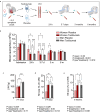
Fig. 4. Neutrophilia, cardiac injury, and response to anti-inflammatory treatment in acute MI are greater in men than women.
ae Data from the double-blind ASSAIL-MI (ASSessing the effect of Anti-IL-6 treatment in MI) study. Patients (167 men, 32 women) with first-time ST-elevation MI were randomized to a single dose of the interleukin (IL)-6 receptor inhibitor tocilizumab or placebo prior to percutaneous coronary intervention (PCI). Men placebo; n = 87, men tocilizumab; n = 80, women placebo; n = 11 and women tocilizumab; n = 21. a Illustration of the study protocol. Created in BioRender. Dahl, T. (2024) https://BioRender.com/p48g864b Blood neutrophil counts during the study. ce Data from cardiac magnetic resonance imaging at 3–7 days (c, d) and 6 months (e) after randomization. LVM left ventricular mass. Bars indicate age-adjusted estimated marginal means, error bars are 95% confidence intervals (CI). Corresponding individual raw data are plotted in Supplementary Fig 9a (for Fig. 4b) and Supplementary Fig 11a–c (for Fig. 4c–e). P values are from two-sided repeated measures ANOVA (b) or ANCOVA (ce) with LSD post-hoc tests. *P < 0.05, **P < 0.01, ***P < 0.001. Exact p values in post-hoc analyses (WP women placebo, MP men placebo, WT women tocilizumab, and MT men tocilizumab); neutrophil count in blood; 24 h; WP vs MP, P = 0.018, WP vs WT, P = 0.002 and MP vs MT, P < 0.001; 3–7 days; WP vs MP, P = 0.025, WP vs WT, P < 0.001 and MP vs MT, P < 0.001; 3 months; WP vs WT, P = 0.019. c LVM; 3–7 days; WP vs MP, P < 0.001 and WT vs MT, P < 0.001. d MI size; 3–7 days; WP vs MP, P = 0.003, WP vs WT, P = 0.035 and MP vs MT, P = 0.012. e MI size; 6 months; WP vs MP, P = 0.016, MP vs MT, P = 0.012.
Fig. 5
Fig. 5. Summary of findings and hypothesis.
In acute MI, the surge in blood neutrophils is a result of neutrophil release from the BM, triggered by various factors, including G-CSF, which suppress the production of neutrophil retention factors, most importantly CXCL12, in the BM. Testosterone (in green) acts via the AR to reduce the production of CXCL12 from BM stromal cells. Homeostatic lower levels of CXCL12 make the BM more prone to release neutrophils in response to egress triggers. The relatively higher blood neutrophils in the presence of male levels of testosterone aggravate myocardial injury in acute MI. Actions of testosterone in men translate into sex differences in neutrophilia and protection by tocilizumab in acute MI-reperfusion. Created in BioRender. Tivesten, Å. (2024) https://BioRender.com/b63s329. G-CSF granulocyte colony-stimulating factor, IL-6 interleukin-6, AR androgen receptor, BM bone marrow.

References

    1. Ng, V. G. et al. Impact of gender on infarct size, ST-segment resolution, myocardial blush and clinical outcomes after primary stenting for acute myocardial infarction: substudy from the EMERALD trial. Int. J. Cardiol.207, 269–276 (2016). - PubMed
    1. Nordlund, D. et al. Gender but not diabetes, hypertension or smoking affects infarct evolution in ST-elevation myocardial infarction patients - data from the CHILL-MI, MITOCARE and SOCCER trials. BMC Cardiovasc. Disord.19, 161 (2019). - PMC - PubMed
    1. De Luca, G. et al. Relation of gender to infarct size in patients with ST-segment elevation myocardial infarction undergoing primary angioplasty. Am. J. Cardiol.111, 936–940 (2013). - PubMed
    1. Berg, J., Bjorck, L., Nielsen, S., Lappas, G. & Rosengren, A. Sex differences in survival after myocardial infarction in Sweden, 1987-2010. Heart103, 1625–1630 (2017). - PMC - PubMed
    1. Cavasin, M. A., Tao, Z., Menon, S. & Yang, X. P. Gender differences in cardiac function during early remodeling after acute myocardial infarction in mice. Life Sci.75, 2181–2192 (2004). - PubMed