. 2025 Feb 6;16(1):1388.
doi: 10.1038/s41467-025-56648-6.

A covalent peptide-based lysosome-targeting protein degradation platform for cancer immunotherapy

Affiliations

A covalent peptide-based lysosome-targeting protein degradation platform for cancer immunotherapy

Youmei Xiao et al. Nat Commun. .

Abstract

The lysosome-targeting chimera (LYTAC) strategy provided a very powerful tool for the degradation of membrane proteins. However, the synthesis of LYTACs, antibody-small molecule conjugates, is challenging. The ability of antibody-based LYTACs to penetrate solid tumor is limited as well, especially to cross the blood-brain barrier (BBB). Here, we propose a covalent chimeric peptide-based targeted degradation platform (Pep-TACs) by introducing a long flexible aryl sulfonyl fluoride group, which allows proximity-enabled cross-linking upon binding with the protein of interest. The Pep-TACs platform facilitates the degradation of target proteins through the mechanism of recycling transferrin receptor (TFRC)-mediated lysosomal targeted endocytosis. Biological experiments demonstrate that covalent Pep-TACs can significantly degrade the expression of PD-L1 on tumor cells, dendritic cells and macrophages, especially under acidic conditions, and markedly enhance the function of T cells and tumor phagocytosis by macrophages. Furthermore, both in anti-PD-1-responsive and -resistant tumor models, the Pep-TACs exert significant anti-tumor immune response. It is noteworthy that Pep-TACs can cross the BBB and prolong the survival of mice with in situ brain tumor. As a proof-of-concept, this study introduces a modular TFRC-based covalent peptide degradation platform for the degradation of membrane protein, and especially for the immunotherapy of brain tumors.


Conflict of interest statement

Competing interests: The authors declare no competing interests.

Figures
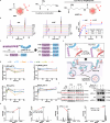
Fig. 1
Fig. 1. Design optimization and functional verification of Pep-TACs.
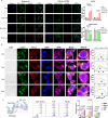
a The schematic diagram of synthesis of covalent amino acids. b HPLC was used to determine the covalent reaction rate of 200 μM k-ASF or ASF with 10 equivalent amounts of Fmoc-D-Lys-OtBu.HCl. n = 3. Data are expressed as means ± SEM. c Sequences of the Pep-TACs. d The effects of DT7, Pep-1 and covalent Pep-TACs on the expression of PD-L1 and TFRC on the surface of B16 and MC38 cells detected by flow cytometry. The P values of PD-L1 level between Pep-1 and v9x across time points (2 h, 6 h, 24 h, 48 h) were <0.001, 0.003, 0.019, 0.026 in B16 cells and 0.024, 0.017, 0.001, 0.203 in MC38 cells, respectively. n = 3. Data are expressed as means ± SEM. e Schematic diagram of the concept and mechanism of covalent Pep-TACs. f The detection of PD-L1 expression in B16 and MC38 cells incubated with covalent Pep-TACs s11x or v9x for 24 or 48 hours by WB. g The detection of the binding of Biotin-v9x (25 μM) to human or mouse PD-L1 protein (2 μg/μL) within a 2-hour incubation at 37 °C by WB. Three independent experiments of WB were performed with similar results and representative results are shown. h The PD-L1 protein (50 μg) and v9x (25 μM) were mixed and incubated at 37 °C for 2 h for the test of protein profiling by intact protein MS. Source data are provided as a Source Data file. All experiments were conducted at least three times and analyzed by unpaired two-tailed t-tests.
Fig. 2
Fig. 2. The endocytosis function of covalent Pep-TACs.
a The endocytosis of 25 μM Biotin-v9x in B16-sh-NT and B16-sh-mTFRC cells (incubated at 37 °C for 2 h) by confocal imaging, scale bar: 20 μm. n = 3. Data are expressed as means ± SEM. b The dynamic colocalization of PD-L1 (red) with lysosomes (Lamp1, green) and TFRC (purple) in B16 cells incubated with 25 μM v9x for 0, 2, 4 or 6 h, as shown by confocal images, scale bar: 5 μm. The white dashed box indicates the magnified region, and the fluorescence gray level plot on the right is derived from the solid white box region. c The affinity between PD-1-Fc protein (100 ng) and PD-L1 on the surface of 293T-mPD-L1 (TFRC low expression), B16, and MC38 cells with 25 μM covalent Pep-TACs for 48 h. n = 3. Data are expressed as means ± SEM. Three independent experiments were performed with similar results and representative results are shown. Source data are provided as a Source Data file. All experiments were analyzed by unpaired two-tailed t-tests.
Fig. 3
Fig. 3. The degradation of PD-L1 by covalent Pep-TAC in evaluated IFN-γ or different pH conditions within tumor, DC or macrophage cell lines.
a The expression of PD-L1 in B16 and MC38 cells incubated with 25 μM v9x in vehicle containing 0.02% DMSO for 24 h at pH7.4 with the presence of 10 μM CQ, or 0.1 μM MG132 by WB. b The expression of PD-L1 in B16 and MC38 cells incubated with 25 μM OPBP1(8-12), Pep-1, or v9x with or without the stimulation of 100 ng/mL IFN-γ for 24 h by flow cytometry. n = 3. Data are expressed as means ± SEM. Statistical differences were determined by two-tailed t-tests. c, d, e Following co-incubation with v9x in vehicle containing 0.02% DMSO at pH6.0 or pH7.4 with or without the treatment of CQ and MG132, the expression of PD-L1 in B16, MC38, DC2.4 or RAW264.7 cells was detected by flow cytometry or WB at indicated times. The P values of PD-L1 level between Pep-1 and v9x at 24 h were 0.025 in B16 cells (pH6.0), 0.005 in MC38 cells (pH6.0), 0.007 (pH7.4) or <0.001 (pH6.0) in DC2.4 cells, 0.212 (pH7.4) or 0.01 (pH6.0) in RAW264.7 cells, respectively. n = 3. Data are expressed as means ± SEM. Source data are provided as a Source Data file. Three independent experiments of WB were performed with similar results and representative results are shown.
Fig. 4
Fig. 4. The in vivo anti-tumor effects of Pep-TACs on MC38 tumor-bearing mice.
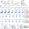
a MC38 tumor model was established and the tumor-bearing mice were treated with indicated drugs as in the schematic diagram. b, c The tumor volume and tumor weight of the tumor-bearing mice, n = 5. Data are expressed as means ± SEM. d, e The representative flow cytometry plots and summary data of the tumor-infiltrating CD8+T cells and IFN-γ+CD8+T cells, n = 4. Data are expressed as means ± SEM. f The representative flow cytometry plots and summary data of the IFN-γ+CD8+T cells in draining lymph node cells by intracellular cytokine staining, n = 4. Data are expressed as means ± SEM. g PD-L1 expression in tumor cells (CD45-), tumor-infiltrating DC (CD45+CD11c+) cells and macrophages (CD45+CD11b+F4/80+), n = 4. Data are expressed as means ± SEM. h The expression of PD-L1 in tumor tissue from MC38 tumor-bearing mice was detected by WB. Three independent experiments were performed with similar results and representative results are shown. Source data are provided as a Source Data file. All experiments were analyzed by unpaired two-tailed t-tests.
Fig. 5
Fig. 5. The in vivo distribution and in vitro BBB transmission of the covalent Pep-TACs.
a The distribution of FITC-GA (2.7 mg/kg, n = 4) and FITC-v9x (5 mg/kg, n = 5) in the tumor-bearing mice was detected by the IVIS system. n = 3. Data are expressed as means ± SEM. b, Pattern of BBB transmission experiments. c HPLC was used to detect the penetration of 50 μM GA, OPBP1(8-12) and v9x cross the BBB, with summary data of the transmission rate of v9x on the right. n = 3. Data are expressed as means ± SEM. Arrows indicate the target peak. d Membrane resistance measurement during BBB permeability experiment. n = 3. Data are expressed as means ± SEM. e The PD-L1 and TFRC expression on the surface of GL261-Luc glioma cells were measured at 2 h, 6 h, 24 h, and 48 h using flow cytometry to determine the effects of 25 μM Pep-1 and v9x. The P values of PD-L1 level between Pep-1 and v9x across time points (2 h, 6 h, 24 h, 48 h) were 0.067, 0.135, 0.261, 0.046, respectively. n = 3. Data are expressed as means ± SEM. Source data are provided as a Source Data file. All experiments were conducted at least three times and analyzed by unpaired two-tailed t-tests.
Fig. 6
Fig. 6. Covalent Pep-TAC v9x inhibits brain tumor growth.
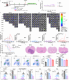
a GL261-Luc brain tumor model was established and the tumor-bearing mice were treated with indicated drugs and the luminescence of the tumors were determined by IVIS and recoded as in the schematic diagram. b The body weight of the tumor-bearing mice during the treatment, Pep-1 and Anti-PD-L1 group: n = 12, Saline and v9x group: n = 13. c, IVIS images of brain tumor in each group. d The change of tumor total flux (p/s) in different groups, Pep-1 and Anti-PD-L1 group: n = 7, Saline and v9x group: n = 8. In parentheses is the number of mice with tumor regression/experimental mice. e, The Kaplan-Meier method with the log rank test was used to examine the survival of tumor-bearing mice. Pep-1 and Anti-PD-L1 group: n = 7, Saline and v9x group: n = 8. f, H&E staining of brain tissue sections after treatment, scale bar: 2.5 mm. Three independent experiments were performed and representative results are shown. g The representative flow cytometry plots and summary data of the tumor-infiltrating T cells, n = 3, meaning that three mice were randomly selected after two weeks of treatment. h The representative flow cytometry plots and summary data of the PD-L1 expression in tumor cells (CD45-), brain microglia (CD45+TMEM119+), tumor-infiltrating DC (CD45+CD11c+) and macrophages (CD45+CD11b+F4/80+), n = 3, meaning that three mice were randomly selected after two weeks of treatment. Source data are provided as a Source Data file. Data are expressed as mean ± standard error (SEM), P values were determined by unpaired two-tailed t-tests.
Fig. 7
Fig. 7. Scheme.
A covalent peptide-based lysosome-targeting protein degradation platform (Pep-TACs) through transferrin receptor (TFRC) for the degradation of membrane proteins, which elicit anti-tumor immune response in both subcutaneous bearing tumors and in situ brain tumors.

References

    1. Schreiber, S. L. The Rise of Molecular Glues. Cell184, 3–9 (2021). - PubMed
    1. Sakamoto, K. M. et al. Protacs: Chimeric molecules that target proteins to the Skp1-Cullin-F box complex for ubiquitination and degradation. Proc. Natl. Acad. Sci. USA98, 8554–8559 (2001). - PMC - PubMed
    1. Nabet, B. et al. The dTAG system for immediate and target-specific protein degradation. Nat. Chem. Biol.14, 431 (2018). - PMC - PubMed
    1. Uhlen, M. et al. Tissue-based map of the human proteome. Science347, 1260419 (2015). - PubMed
    1. Banik, S. M. et al. Lysosome-targeting chimaeras for degradation of extracellular proteins. Nature584, 291 (2020). - PMC - PubMed

MeSH terms