

CX3CR1+ age-associated CD4+ T cells contribute to synovial inflammation in late-onset rheumatoid arthritis
- PMID: 39910629
- PMCID: PMC11800492
- DOI: 10.1186/s41232-025-00367-4
CX3CR1+ age-associated CD4+ T cells contribute to synovial inflammation in late-onset rheumatoid arthritis
Abstract
Background: Recent evidence suggests that clonally expanded cytotoxic T cells play a role in various autoimmune diseases. Late-onset rheumatoid arthritis (LORA) exhibits unique characteristics compared to other RA forms, suggesting distinct immunological mechanisms. This study aimed to examine the involvement of cytotoxic T cells in LORA.
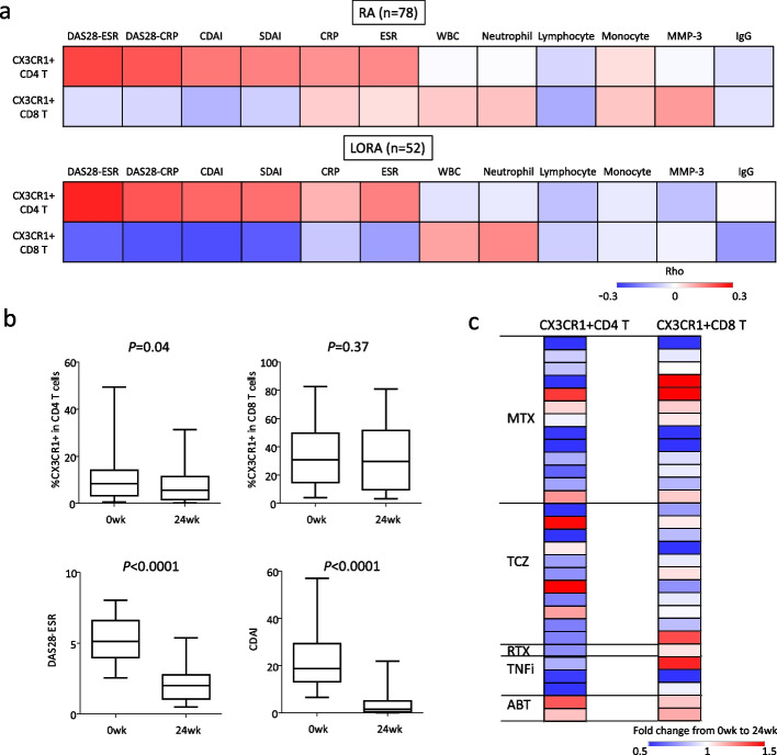
Methods: Fresh peripheral blood samples were collected from 78 treatment-naïve active RA patients, 12 with difficult-to-treat RA, and 16 healthy controls. Flow cytometry was employed to measure the proportions of CX3CR1+cytotoxic CD4+ and CD8+ T cells in these samples. Additionally, immunohistochemical staining was performed on lymphoid node and synovial biopsy samples from patients with RA.
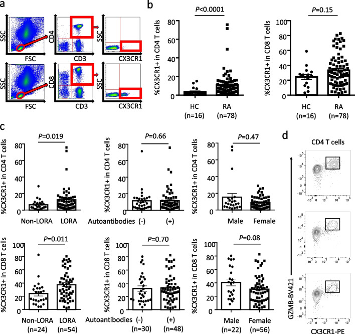
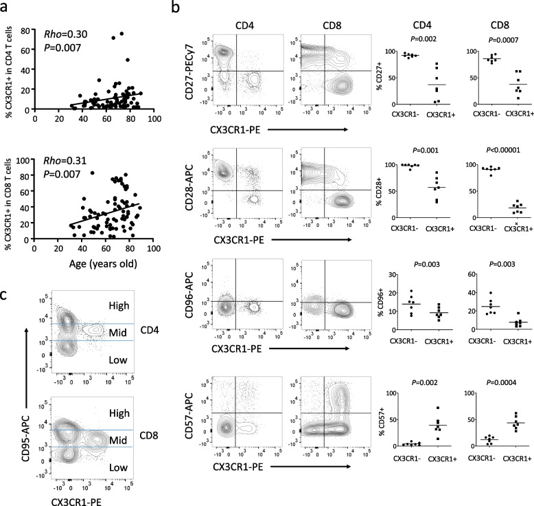
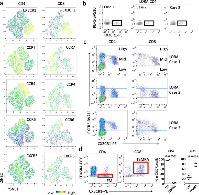
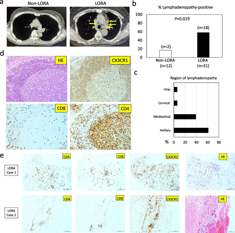
Results: CX3CR1+cytotoxic CD4+ T cells were specifically increased in untreated, active patients with LORA, displaying features of CXCR3mid age-associated T helper cells known as "ThA". CX3CR1⁺CD4⁺ T cells were identified as a cytotoxic ThA subset, as nearly all of these cells specifically expressed granzyme B. These cells were observed in enlarged lymph nodes and were found to infiltrate synovial tissues from patients with LORA. The proportions of CX3CR1+CD4+ T cells positively correlated with arthritis activity in LORA. The number of cells decreased after treatment with methotrexate, tumor necrosis factor inhibitors, and interleukin-6 inhibitors, whereas T-cell activation modulators did not affect them. Moreover, PD-1+CD38+CX3CR1+CD4+ T cells were identified as a treatment-resistant T cell subset that was characteristically increased in difficult-to-treat RA. CX3CR1+CD8+ T cells showed no significant difference between RA patients and healthy individuals, and no correlation with disease activity was observed. However, a correlation with age was observed in RA patients.
Conclusions: Our findings suggest that the immunopathogenesis of RA differs by age of onset, with CX3CR1+ age-associated cytotoxic CD4+ T cells playing a significant role in LORA. Additionally, the presence of a specific CX3CR1+ T cell subset may be linked to treatment resistance.
Keywords: Aging; CX3CR1; Cytotoxic T cells; Difficult-to-treat rheumatoid arthritis; Late-onset rheumatoid arthritis; Rheumatoid arthritis.
© 2025. The Author(s).
Conflict of interest statement
Declarations. Ethics approval and consent to participate: Approval for the study was granted by the Ethics Committee of Keio University School of Medicine (Approval number: 20140335). The study was conducted in accordance with the principles outlined in the Declaration of Helsinki. Written informed consent was obtained from all the participants. Consent for publication: Not applicable. Competing interests: M.A. has received speaking fees from AbbVie, Asahi-Kasei, Astellas, AstraZeneca, Boehringer Ingelheim, Bristol-Myers Squibb, Chugai, Eisai, Eli Lilly, Janssen, Novartis, Pfizer, Sanofi, Taisho, UCB, Gilead Sciences, Glaxo Smith Kline. Y.Ko. has received speaking fees from AbbVie, Ayumi Pharmaceutical Corporation, Boehringer Ingelheim, Bristol-Myers Squibb, Chugai, Eisai, Eli Lilly, Glaxo Smith Kline, Gilead Sciences, Pfizer and Taisyo. Y. Ka. has received research grants from AbbVie, Eisai, Sanofi, Chugai, Mitsubishi-Tanabe, Taisho, and has received scholarship grant from Asahi-Kasei, Eisai, Boehringer Ingelheim, Taisho, and has received speaking fees from AbbVie, Asahi-Kasei, Astellas, Ayumi Pharmaceutical Corporation, Boehringer Ingelheim, Bristol-Myers Squibb, Chugai, Eisai, Eli Lilly, Glaxo Smith Kline, Janssen, Novartis, Pfizer, UCB, Gilead Sciences. S. W., K.Y., K.S., S.I., R.I., Y.M., W.A. declare no conflict of interest.
Figures

References
-
- McInnes IB, Schett G. The pathogenesis of rheumatoid arthritis. N Engl J Med. 2011;365:2205–19. - PubMed
-
- Gravallese EM, Firestein GS. Rheumatoid Arthritis - Common Origins. Divergent Mechanisms N Engl J Med. 2023;388:529–42. - PubMed
-
- Malmström V, Catrina AI, Klareskog L. The immunopathogenesis of seropositive rheumatoid arthritis: from triggering to targeting. Nat Rev Immunol. 2017;17:60–75. - PubMed
-
- Smolen JS, Aletaha D, Barton A, Burmester GR, Emery P, Firestein GS, et al. Rheumatoid arthritis Nat Rev Dis Primers. 2018;4:18001. - PubMed
LinkOut - more resources
Full Text Sources
Research Materials
