. 2025 Feb;15(2):e70239.
doi: 10.1002/brb3.70239.

Aldh1l1-Cre/ERT2 Drives Flox-Mediated Recombination in Peripheral and CNS Infiltrating Immune Cells in Addition to Astrocytes During CNS Autoimmune Disease

Affiliations

Aldh1l1-Cre/ERT2 Drives Flox-Mediated Recombination in Peripheral and CNS Infiltrating Immune Cells in Addition to Astrocytes During CNS Autoimmune Disease

Mario Amatruda et al. Brain Behav. 2025 Feb.

Abstract

Introduction: The transgenic murine Cre/loxP system is deployed to investigate the role of central nervous system (CNS) cell-specific gene alterations in both healthy conditions and models of neurologic disease. The Aldh1l1-Cre/ERT2 line is widely used to target astrocytes with high coverage and specificity within the CNS. Specificity outside the CNS, however, has not been well-characterized, and Aldh1l1-Cre/ERT2-mediated recombination within the spleen has been reported. In many CNS diseases, infiltrating immune cells from the periphery drive or regulate pathogenesis. We tested whether flox-mediated recombination from Aldh1l1-Cre/ERT2 occurs in immune cells in addition to astrocytes and whether these cells traffic from the spleen into the spinal cord during experimental autoimmune encephalomyelitis (EAE), a model of CNS autoimmune disease.

Methods: Two astrocyte-targeted mouse lines were generated with the red fluorescent reporter, tdTomato, by crossing the Cre-recombinase lines, Tg(Aldh1l1-Cre/ERT2)1Khakh and Tg(Gfap-Cre)73.12Mvs, with the reporter line, Gt(ROSA)26Sor. Aldh1l1-Cre/ERT2 was activated with 5 days of intraperitoneal tamoxifen, whereas Gfap-Cre was constitutively active. EAE was induced 2 weeks after tamoxifen, and then spleens and spinal cords were harvested and processed for flow cytometry at various time points after disease onset in EAE versus healthy controls.

Results: In EAE, Aldh1l1-Cre/ERT2, but not Gfap-Cre, induced multiple tdTomato+ immune cell subpopulations in the spleen and spinal cord, including macrophages, monocytes, neutrophils, eosinophils, B cells, CD4+, and CD8+ T cells.

Conclusion: Use of Aldh1l1-Cre/ERT2 should therefore account for recombination in both astrocytes and immune cells in disease models involving peripheral immune cell infiltration into the CNS.

Keywords: Aldh1l1‐Cre; CNS infiltrating immune cell; Gfap‐Cre; astrocyte‐specific promoter; central nervous system (CNS) autoimmune disease; experimental autoimmune encephalomyelitis (EAE); flow cytometry.


Conflict of interest statement

The authors declare no conflicts of interest. Dr. Mario Amatruda was previously employed as a senior scientist at Biogen in 2023.

Figures

FIGURE 1
FIGURE 1
Aldh1l1‐Cre/ERT2;ROSA mice with EAE are characterized by the presence of CD45+/tdTomato+ cells in the inflamed spinal cord. Spinal cords from mice with EAE and under healthy conditions were harvested 6 weeks after treatment with tamoxifen (at 28 days post EAE immunization), homogenized and centrifuged in a 70%/30% Percoll gradient to enrich the astrocytic cell population. DNA recombination was assessed following the expression of the tdTomato reporter gene in flow cytometry. (A) As expected, no tdTomato+ cells were observed in ROSA control mice (with no Aldh1l1‐Cre/ERT2). tdTomato+ cells were detected in Aldh1l1‐Cre/ERT2;ROSA mice, indicating successful DNA recombination. Aldh1l1‐Cre/ERT2; ROSA mice with EAE were characterized by the presence of an additional distinctive population of cells positive for tdTomato (red rectangle) which was absent in Aldh1l1‐Cre/ERT2;ROSA healthy controls. (B) Flow cytometry analysis revealed that this distinct tdTomato+ cell population was positive for the leukocyte common antigen, CD45. EAE, experimental autoimmune encephalomyelitis.
FIGURE 2
FIGURE 2
Aldh1l1‐Cre/ERT2 drives loxP recombination in CD45+ leukocytes. Spleens were harvested from Aldh1l1‐Cre/ERT2;ROSA mice with EAE and from healthy controls (HC) 6 weeks after treatment with tamoxifen. DNA recombination was assessed following the expression of the tdTomato reporter gene in flow cytometry. (A and B) Expression of the tdTomato reporter gene was observed in the spleen of both HC (range: 33.8%–45.4% of total live cells) and EAE (range: 31.3%–37.9%) mice. The proportion of cells positive for tdTomato over the total number of live cells was not significantly different between HC and EAE mice. (C and D) Almost all tdTomato+ splenocytes were CD45+, indicating that the Aldh1l1 promoter drives loxP recombination in leukocytes. Mice with EAE and HCs showed similar proportions of tdTomato+ CD45+ cells over the total number of tdTomato+ cells (HC, mean = 98.2; EAE, mean = 98.9). (E and F) tdTomato+ cells represented about 40% of the total live CD45+ cells in the spleen in both HC (range: 44.3%–48.8%) and EAE (range: 34.4%–42.5%) mice. No statistically significant differences were observed between the two groups. (B, D, and F) n = 3 per group; unpaired Student's t test. EAE, experimental autoimmune encephalomyelitis.
FIGURE 3
FIGURE 3
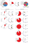
Characterization of CD45+ tdTomato+ cells in Aldh1l1‐Cre/ERT2 mice. Flow cytometry of the CD45+ tdTomato+ cells from the spleen of Aldh1l1‐Cre/ERT2 mice 3 weeks after tamoxifen treatment. Pie charts show averaged cell populations’ frequency (n = 3 mice), whereas density plots show representative FACS analysis per each cell population. (A) Pie chart shows the relative frequency of the analyzed leukocyte cell types over the total number of CD45+ cells in spleen. The antibody panel used in this experiment did not resolve 15.3% of the CD45+ cells. (B) On average, 21% of CD45+ cells were tdTomato+. (C) Pie charts show the proportion of each leukocyte subset over the total number of CD45+ tdTomato+ cells. (D and E) Pie charts show the average proportion of tdTomato+ cells in each specific cell type from the lymphoid (D) and myeloid (E) lineage. Representative density plots show the frequency of tdTomato+ cells within each leukocyte subtype. Gating strategies are depicted in Figures S1 and S2.
FIGURE 4
FIGURE 4
Gfap‐Cre does not significantly drive loxP recombination in splenic leukocytes. tdTomato reporter expression in Aldh1l1‐Cre/ERT2;ROSA 3 weeks after treatment with tamoxifen and untreated Gfap‐Cre;ROSA mice (A–D). ROSA mice (with no Cre) were used as controls. (A and B) Flow cytometry (representative density plots in A; quantification in B) analysis shows a significant greater proportion of tdTomato+ CD45+/CD45+ leukocytes in the spleen of Aldh1l1‐Cre/ERT2;ROSA (mean 19.4) compared with Gfap‐Cre;ROSA (mean 2.5) and control mice (mean 0.94), in which tdTomato+ cells were nearly undetectable, Aldh1l1‐Cre/ERT2;ROSA versus Gfap‐Cre;ROSA p < 0.005, Aldh1l1‐Cre/ERT2;ROSA versus ROSA, p < 0.005, Gfap‐Cre;ROSA versus ROSA, p = 0.83; n = 3 per group. One‐way ANOVA with Tukey's multiple comparison test. (C and D) RT‐PCR analysis of tdTomato transcript levels revealed a significant upregulation in the spinal cord of both Aldh1l1‐Cre/ERT2;ROSA and Gfap‐Cre;ROSA compared with controls (C) (means 21.98, 15.92, and 1.0, respectively; Aldh1l1‐Cre/ERT2;ROSA vs. Gfap‐Cre;ROSA, p < 0.05, Aldh1l1‐Cre/ERT2;ROSA vs. ROSA, p < 0.0001, Gfap‐Cre;ROSA vs. ROSA, p < 0.0005). n = 3 per group. One‐way ANOVA with Tukey's multiple comparison test. tdTomato transcript levels were significantly increased in the spleen of Aldh1l1‐Cre/ERT2;ROSA mice compared with Gfap‐Cre;ROSA mice and ROSA controls (D) (means 6.79, 1.33, and 1.0, respectively; Aldh1l1‐Cre/ERT2;ROSA vs. Gfap‐Cre;ROSA, p < 0.0005, Aldh1l1‐Cre/ERT2;ROSA vs. ROSA, p < 0.0005, Gfap‐Cre;ROSA vs. ROSA, p = 0.87), suggesting non‐astrocyte specific loxP recombination in Aldh1l1‐Cre/ERT2 mice but not in Gfap‐Cre mice. n = 3 per group. One‐way ANOVA with Tukey's multiple comparison test. (E and F) Immunohistochemistry images (E) and co‐localization analysis (F) of tdTomato and CD45 staining in spleens of healthy, tamoxifen‐treated Aldh1l1‐Cre/ERT2;ROSA and Gfap‐Cre;ROSA mice confirmed the presence and significantly increased levels of co‐localizing tdTomato+ (red) and CD45+ (green) signal in Aldh1l1‐Cre/ERT2;ROSA mice compared to Gfap‐Cre;ROSA mice (mean 0.62 vs. 0.24, respectively, p < 0.001). Samples from both groups were harvested 2 weeks post‐tamoxifen treatment. Scale bar 50 µm, n = 3 per group. Unpaired Student's t‐test.
FIGURE 5
FIGURE 5
Representative FACS density plots for tdTomato+ leukocytes from EAE spinal cords of Aldh1l1‐Cre/ERT2;ROSA and Gfap‐Cre;ROSA mice at peak disease. Aldh1l1‐Cre/ERT2;ROSA mice show tdTomato+ populations of neutrophils, macrophages, monocytes, and CD4+ T cells, whereas Gfap‐Cre;ROSA mice lack these populations. Gating strategies are depicted in Figures S3 and S4.

References

    1. Absinta, M. , Maric D., Gharagozloo M., et al. 2021. “A Lymphocyte‐Microglia‐Astrocyte Axis in Chronic Active Multiple Sclerosis.” Nature 597, no. 7878: 709–714. 10.1038/s41586-021-03892-7. - DOI - PMC - PubMed
    1. Amatruda, M. , Chapouly C., Woo V., et al. 2022. “Astrocytic Junctional Adhesion Molecule‐A Regulates T‐Cell Entry Past the Glia Limitans to Promote Central Nervous System Autoimmune Attack.” Brain Communications 4, no. 2: fcac044. 10.1093/braincomms/fcac044. - DOI - PMC - PubMed
    1. Batiuk, M. Y. , Martirosyan A., Wahis J., et al. 2020. “Identification of Region‐Specific Astrocyte Subtypes at Single Cell Resolution.” Nature Communications 11, no. 1: 1220. 10.1038/s41467-019-14198-8. - DOI - PMC - PubMed
    1. Benallegue, N. , Kebir H., Kapoor R., et al. 2021. “The Hedgehog Pathway Suppresses Neuropathogenesis in CD4 T Cell‐Driven Inflammation.” Brain 144, no. 6: 1670–1683. 10.1093/brain/awab083. - DOI - PMC - PubMed
    1. Beyer, F. , Ludje W., Karpf J., Saher G., and Beckervordersandforth R.. 2021. “Distribution of Aldh1L1‐CreER(T2) Recombination in Astrocytes Versus Neural Stem Cells in the Neurogenic Niches of the Adult Mouse Brain.” Frontiers in Neuroscience 15: 713077. 10.3389/fnins.2021.713077. - DOI - PMC - PubMed

MeSH terms

LinkOut - more resources