Review
. 2025;3(1):4.
doi: 10.1038/s44324-025-00049-2. Epub 2025 Feb 3.

Calcium-mediated regulation of mitophagy: implications in neurodegenerative diseases

Affiliations
Review

Calcium-mediated regulation of mitophagy: implications in neurodegenerative diseases

Fivos Borbolis et al. NPJ Metab Health Dis. 2025.

Abstract

Calcium signaling plays a pivotal role in diverse cellular processes through precise spatiotemporal regulation and interaction with effector proteins across distinct subcellular compartments. Mitochondria, in particular, act as central hubs for calcium buffering, orchestrating energy production, redox balance and apoptotic signaling, among others. While controlled mitochondrial calcium uptake supports ATP synthesis and metabolic regulation, excessive accumulation can trigger oxidative stress, mitochondrial membrane permeabilization, and cell death. Emerging findings underscore the intricate interplay between calcium homeostasis and mitophagy, a selective type of autophagy for mitochondria elimination. Although the literature is still emerging, this review delves into the bidirectional relationship between calcium signaling and mitophagy pathways, providing compelling mechanistic insights. Furthermore, we discuss how disruptions in calcium homeostasis impair mitophagy, contributing to mitochondrial dysfunction and the pathogenesis of common neurodegenerative diseases.

Keywords: Metabolic disorders; Mitochondria.


Conflict of interest statement

Competing interestsThe authors declare no competing interests.

Figures

Fig. 1
Fig. 1. Intracellular Ca2+ trafficking.
Ca2+ ions are constantly transferred from the cytoplasm to the extracellular space, through the action of the plasma membrane Ca2+ ATPase PMCA and Na+/Ca2+ (NCX) or Na+/Ca2+-K+ (NCKXs) exchangers. Opening of voltage-gated or ligand-gated plasma membrane Ca2+ channels (VGCCs or LGCCs) results in the rapid influx of Ca2+ ions along their chemical gradient. Inside the cell, large amounts of Ca2+ are transferred in the ER by the sarcoendoplasmic reticulum Ca2+-ATPase (SERCA) and can be released upon stimulation via IP3R or RyR channels. Depletion of ER Ca2+ stores induces the translocation of STIM protein to ER/PM contact sites where it activates the Ca2+ channel Orai to mediate prolonged influx of extracellular Ca2+. Ca2+ is also stored in the cis-Golgi through its transfer by SERCA or the trans-Golgi via the Ca2+-ATPase SPCA and can be released through IP3R or RyR channels, respectively. The Ca2+ content of lysosomes, built up by the transfer of ions from the cytoplasm via an unidentified importer, can be released through the action of transient receptor potential mucolipin channels (TRPML) and two-pore channels (TPC). The OMM is easily penetrated by Ca2+ ions that pass through VDAC but has to be transferred by the highly selective mitochondrial Ca2+ uniporter (mtCU) complex of the IMM in order to reach the matrix. Ca2+ export from mitochondria is mediated by the Na+/Ca2+ exchanger NCLX and the Ca2+/H+ antiporter Leucine Zipper And EF-Hand Containing Transmembrane Protein 1 (LETM1). Ca2+ overload or dissipation of Δψm can induce the opening of the mitochondrial permeability transition pore (mPTP), which mediates the rapid efflux of Ca2+ to the cytoplasm. Depolarization (shown in red) or altered pH (shown in yellow) can allow the reversal of NCLX or LETM1, respectively, transforming them into Ca2+ importers that function with different properties than the mtCU. For details see text. Created in BioRender. Palikaras, K. (2024) https://BioRender.com/v71t847.
Fig. 2
Fig. 2. Interplay between calcium homeostasis and mitophagy.
A At low cytosolic calcium levels, Miro1 facilitates mitochondrial movement by interacting with Milton/TRAK. Increased Ca2+ disrupts this interaction, exposing Miro1 to ubiquitination by Parkin and the subsequent PINK1/Parkin-dependent mitophagy upon concurrent mitochondrial stress. B CCCP-induced mitochondrial depolarization triggers the demultimerization of Miro2 from a tetramer to a monomer, a process dependent on PINK1 phosphorylation and Ca2+ binding to Miro2 EF2 domain. Miro2 re-alignment stimulates Parkin translocation and mitophagy initiation. C Cytosolic calcium accumulation activates the kinase CAMKIα and the phosphatase CaN which mediate the phosphorylation (Ser600) and dephosphorylation (Ser637) of DRP1, respectively. Both modifications promote mitochondrial translocation of DRP1 which triggers fission, priming mitochondria for mitophagic degradation. D In pancreatic β cells, mitochondrial stress generates ROS that act as signaling molecules for lysosomal calcium release via TRPML channels. Elevated Ca2+ activates CaN, which, in turn, dephosphorylates TFEB, enabling its nuclear translocation. In the nucleus, TFEB promotes the expression of the mitophagy adaptor proteins NDP52 and OPTN, among others. Lysosomal calcium stores are primarily replenished from ER. E The key mitophagy factor PINK1 facilitates mitochondrial calcium release by phosphorylating NCLX and LETM1 exchangers, thereby preventing Ca2+ overload within mitochondria. PINK1 depletion results in mitochondrial calcium overload, increased ROS generation, mPTP opening and a subsequent rise in cytosolic Ca2+ due to diminished mitochondrial buffering capacity. F Examples of Parkin implication in the regulation of calcium homeostasis. Parkin prevents cytosolic calcium overload by ubiquitinating and promoting the proteasomal degradation of PLCγ, the upstream activator of IP3R. CISD1 has been identified as the downstream effector for Parkin-dependent regulation of ER-calcium release. Parkin is proposed to initially enhance ER-mitochondria tethering and mitochondrial Ca2+ influx as a means to boost bioenergetics (dashed green arrows). Failure in restoring bioenergetic imbalance enables Parkin’s mitophagy-inducing activity for the elimination of compromised mitochondria (dashed red arrow). Moreover, the cytosolic fraction of Parkin impedes mitochondrial calcium uptake by interacting with MICU1 precursor, enhancing its ubiquitination and subsequent proteasomal degradation. Created in BioRender. Palikaras, K. (2024) https://BioRender.com/r42n942.
Fig. 3
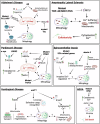
Fig. 3. Links between Ca2+ homeostasis and mitophagy in the context of neurodegenerative disorders.
Dashed lines indicate putative links. Continuous lines correspond to links supported by experimental evidence. For details see “Calcium dysregulation and mitophagy in neurodegeneration.” Created in BioRender. Borbolis, F. (2024) https://BioRender.com/e98r825.

References

    1. Bootman, M. D. & Bultynck, G. Fundamentals of cellular calcium signaling: a primer. Cold Spring Harb. Perspect. Biol.12, a038802 (2020). - PMC - PubMed
    1. Giorgi, C., Marchi, S. & Pinton, P. The machineries, regulation and cellular functions of mitochondrial calcium. Nat. Rev. Mol. Cell Biol.19, 713–730 (2018). - PubMed
    1. Berridge, M. J., Lipp, P. & Bootman, M. D. The versatility and universality of calcium signalling. Nat. Rev. Mol. Cell Biol.1, 11–21 (2000). - PubMed
    1. Bagur, R. & Hajnóczky, G. Intracellular Ca2+ sensing: its role in calcium homeostasis and signaling. Mol. Cell66, 780–788 (2017). - PMC - PubMed
    1. Rossi, A., Pizzo, P. & Filadi, R. Calcium, mitochondria and cell metabolism: a functional triangle in bioenergetics. Biochim. Biophys. Acta Mol. Cell Res.1866, 1068–1078 (2019). - PubMed

LinkOut - more resources