. 2025 Jan 16:7:100142.
doi: 10.1016/j.bbadva.2025.100142. eCollection 2025.

New insights into the N-glycomes of Dictyostelium species

Affiliations

New insights into the N-glycomes of Dictyostelium species

Alba Hykollari et al. BBA Adv. .

Abstract

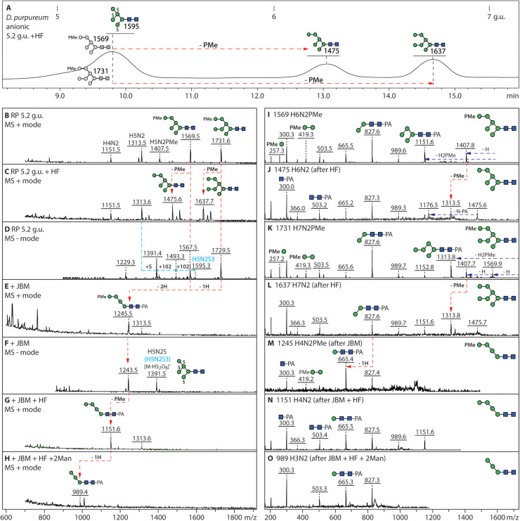
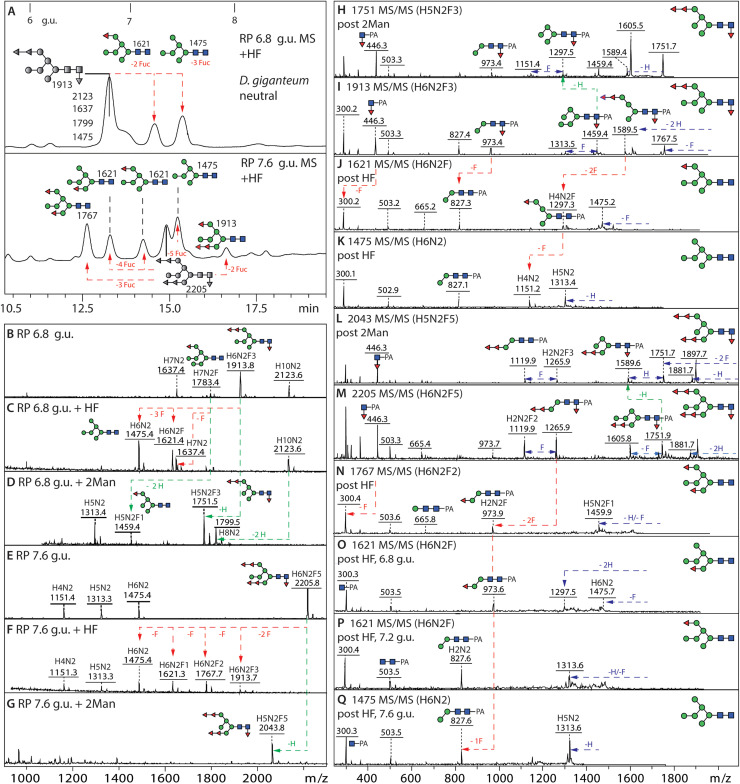
Dictyostelia are cellular slime molds, a group of Amoebozoa, that form multicellular fruiting bodies out of aggregating cells able of differentiating into resistant spore forms. In previous studies on Dictyostelium discoideum, it was demonstrated that their N-glycans, as in most eukaryotes, derive from the Glc3Man9GlcNAc2-PP-Dol precursor; however, unique glyco-epitopes, including intersecting GlcNAc, core α1,3-fucosylation, sulphation and methylphosphorylation, were detected. In the present study, we have examined the N-glycans of two other Dictyostelium species, D. purpureum, whose genome is also sequenced, and D. giganteum. The detailed glycomic analysis of their fruiting bodies was based on isomeric separation of the glycan structures by HPLC, followed by mass spectrometry in combination with enzymatic digests and chemical treatments. Two features absent from the 'model' dictyostelid D. discoideum were found: especially in D. purpureum, a long linear galactose arm β1,4-linked to the β1,4-N-acetylglucosamine on the 'lower' A-branch of its oligo-mannosylated structures could be identified. In contrast, neutral N-glycans with multiple fucose residues attached to terminal mannoses were found in D. giganteum. All three species have common modifications on their anionic N-glycans: while (methyl)phosphorylated residues are always associated with terminal mannose residues, the sulphation position differs. While D. discoideum has 6-sulphation of subterminal mannose residues, D. giganteum and D. purpureum may rather have 2-sulphation of core α1,6-mannose. Overall, we have discovered species-specific glycan variations and our data will contribute to future comparative and functional studies on these three species within the same genus.

Keywords: Dictyostelia; Fucose; Galactose; N-glycan; Phosphate; Sulphate.


Conflict of interest statement

The authors declare the following financial interests/personal relationships which may be considered as potential competing interests: Katharina Paschinger reports financial support was provided by Austrian Science Fund. If there are other authors, they declare that they have no known competing financial interests or personal relationships that could have appeared to influence the work reported in this paper.

Figures

Fig. 1
Fig. 1
RP-HPLC analysis of pyridylaminated N-glycans from D. purpureum. (A and B) RP-amide HPLC chromatograms of pyridylaminated neutral and anionic PNGase F and A-released N-glycans from the fruiting bodies of D. purpureum. Enrichment by NPGC-SPE resulted in neutral and anionic pools prior to pyridylamination (see Supplementary Figure 1 for the overall MALDI-TOF MS) and RP-HPLC. The reverse phase column was calibrated in terms of glucose units (g.u.). Structures are annotated according to the nomenclature of the Symbolic Nomenclature for Glycomics (circles: mannose in green and galactose in yellow; squares: N-acetylglucosamine in blue; triangle: fucose in red; S suphate; P phosphate; Me methyl groups; see inset) on the basis of retention time and MS/MS with or without chemical/enzymatic treatments; m/z values are shown as [M + H]+ or in italics, for sulphated glycans, [M-H]- or [M-nH+(n-1)Na]-. Sulphated structures are detected only in negative mode, whereby monosulphated glycans were detected as [M-H]- and di-, tri- and tetrasulphated ones in sodiated form; in positive mode, the sulphate is lost in source (m/z values in square brackets). Structures common to both D. purpureum and D. giganteum are highlighted with grey squares; structures are shown in accordance with the observed abundance in the individual fractions (most abundant uppermost). Oligomannosidic glycans dominate the neutral pool; in addition, there are early-eluting core α1,3-fucosylated Hex1–7HexNAc2Fuc1 and late eluting Hex3–12HexNAc3 glycans. The anionic pool contains many of the same oligomannosidic-type glycans modified mainly with methylphosphate or sulphate, but lacks galactosylated forms. The terms A-arm, B-arm, C-arm and intersect are defined for an example Hex8HexNAc3 glycan.
Fig. 2
Fig. 2
RP-HPLC fractionation of neutral and anionic pyridylaminated N-glycans fromD. giganteum. (A and B) RP-amide HPLC chromatograms of pyridylaminated neutral and anionic PNGase F and A-released N-glycans from the fruiting bodies of D. giganteum (see Supplementary Figure 1 for the overall MALDI-TOF MS). As in Fig. 1, the chromatogram is annotated with glucose units and each glycan is shown as a symbolic structure (see also inset) together with the relevant m/z value. Structures common to both D. purpureum and D. giganteum are highlighted with grey squares, especially including oligomannosidic and monofucosylated glycans. In contrast to D. discoideum and D. purpureum, more glycans are core fucosylated with up to six further fucose residues substituting terminal mannoses. The anionic pool contains many of these fucosylated glycans modified with methylphosphate or sulphate, but the presence of antennal fucose correlates with a lower degree of sulphation, i.e., only three multisulphated structures as compared to seventeen in D. purpureum. The oligomannosidic methylphosphorylated structures are shared in both species, but multifucosylated monosulphated ones occur only in D. giganteum.
Fig. 3
Fig. 3
Phospho-mono- and diesters as modifications of mannose residues of D. purpureum anionic N-glycans. (A, C) The 4.3 g.u. fraction of the D. purpureum anionic pool was subject to positive and negative mode MALDI-TOF MS showing various phosphorylated, methylphosphorylated and phosphodiester-modified structures. (B) Hydrofluoric acid treatment resulted in loss of the phosphoester modifications. (D-F) Jack bean α-mannosidase digestion resulted in products of Man4–5GlcNAc2–3P, converted subsequently to Man4–5GlcNAc2 upon HF treatment and the combined treatment caused a change in RP-amide elution time correlating with the annotated isomer; alterations are shown by red arrows. (G-I) Positive and negative mode MS/MS of the phosphodiester modified Man7GlcNAc3P structure shows the GlcNAc-1-P of the “upper” C arm, whereby the negative ion mode m/z 444 and 1416 B1 and B4 fragments (H) or the positive mode loss of GlcNAc1P1Man2 (607 Da) are indicative of the HexNAcPMan modification; MS/MS of the product of HF treatment shows the underlying Man7GlcNAc2 isomer. (J-L) MALDI-TOF MS/MS of two glycans modified with unmodified phosphate and the α-mannosidase product. (M) MALDI-TOF MS/MS of a core fucosylated structure in the same fraction modified with methylphosphate. Further examples of phosphoester-modified glycans are shown in Supplementary Figures 3 and 4.
Fig. 4
Fig. 4
Combined enzymatic, chemical and RP HPLC approach for definition of the methylphosphate modification. (A-D) The 5.2. g.u. RP HPLC fraction of the D. purpureum anionic N-glycan pool contained two glycans with single methylphosphate modifications, which were removed by HF treatment resulting in a shift in retention time and mass, thereby definining the underlying oligomannosidic isomers. (E-H) In parallel, jack bean mannosidase digestion resulted in a major Man4GlcNAc2PMe, indicating that the C-arm was blocked by the methylphosphate moiety, which was then removed by subsequent HF treatment; finally, the dephosphorylated glycan was sensitive to α1,2-mannosidase, resulting in an α1,6-mannosidase-sensitive product of m/z 989. Note that the co-eluting m/z 1595 glycan (D) is insensitive to α-mannosidase digestion due to the three sulphate residues, which are labile during mass spectrometry, but resistant to HF. (I-O) Positive mode MALDI-TOF MS of the major methylphosphorylated glycans in the 5.2. g.u. fraction and their digestion products showing shifts in the pattern of B and Y-ions. Further examples of methylphosphorylated glycans are shown in Supplementary Figures 3, 4 and 5.
Fig. 5
Fig. 5
MALDI TOF MS and LC-MSn analysis of the galactose modified N-glycan in D. purpureum and D. giganteum. Analysis of two isomeric N-glycan structures, separated by RP HPLC of the D. purpureum neutral N-glycan pool at 16.2 and 17.5 g.u (m/z 2002; Hex8HexNAc3) in positive ion mode with MALDI TOF MS/MS before and after exoglycosidase treatment. (A-F) Jack bean α-mannosidase or β1,4-specific galactosidase and subsequent chitinase (β1,3/4-hexosaminidase; see inset) treatment of the major isomer at 16.2 g.u. resulted in products of m/z 1516 and 1313 and shifts in the MS/MS spectra, as indicated with red arrows, indicative of a trigalactose motif on the lower arm, which correlated with the m/z 690 B-fragment; this glycan was insensitive to α1,2-mannosidase treatment (not shown). (G) Negative mode LC-MSn of the major D. purpureum trigalactosylated Hex8HexNAc3 glycan shows antennal 0.2A-Gal and 2,4A-GlcNAc fragments compatible with a β1,4-linkage of the galactose residues. (H, I) The later eluting m/z 2002 isomer (17.5 g.u.) otherwise showing the same key m/z 690 and 1313 fragments, lost one hexose residue after α1,2-mannosidase treatment, indicative of a different underlying Man5GlcNAc2 backbone. (J, K) The galactosylated epitopes were also identified on some glycans from D. giganteum, whereby these structures were generally core α1,3-fucosylated as defined by the m/z 446 Y1-fragment ions, while m/z 528 and 690 B-fragments correlated with di- or trigalactose motifs on the “lower” A arm. Properties of the glycans with underlying non-reducing terminal GlcNAc are shown in Supplementary Figure 6 and further MS/MS of isomers with mono- up to penta-galactosylated epitopes are presented in Supplementary Figure 7.
Fig. 6
Fig. 6
RP-HPLC and MSMS analysis of N-glycans from D. giganteum modified with both core and terminal fucose. (A) RP-Amide chromatograms of the two hydrofluoric acid (HF) treated 6.8 and 7.8 g.u. fractions of the neutral D. giganteum N-glycan pool showing shifts in retention time due to serial losses of fucose, revealing the underlying Man6GlcNAc2 structure. (B-G) MALDI-TOF MS of these fractions shows that the fucose residues, core linked to the reducing terminal N-acetylglucosamine residue and/or terminally linked to mannose were sensitive to HF treatment (red arrows), while specific α1,2-mannosidase treatment resulted in loss of one or two hexose residues (green arrows). (H-Q) Positive mode MALDI TOF MS/MS of the untreated, HF-treated and mannosidase-treated glycans highlights changes in the fragmentation patterns due to serial removal of fucose or removal of mannose. While the Y3-fragment at m/z 1265 (Hex2HexNAc2Fuc3-PA) is indicative of a fucosylated A arm, a fragment at m/z 973 (Hex2HexNAc2Fuc1-PA) can be either core or antennally fucosylated; a Y1 fragment at m/z 446 shows core fucosylation (see also Supplementary Figure 8). The final defucosylation products have Y3 fragments at m/z 827 (Hex2HexNAc2-PA). Note that the core α1,3-fucose was more sensitive than the antennal ones to HF treatment; for further multi-fucosylated isomers, refer to Supplementary Figure 9. Furthermore, the antennal fucose residues, but not the core α1,3-fucose, were partially released by bovine α-fucosidase, while all fucose residues were resistant to microbial α1,2-fucosidase and almond α1,3/4-fucosidase (data not shown).
Fig. 7
Fig. 7
Multiple sulphation in D. purpureum and fucosylated sulphated structures in D. giganteum. (A-F) Negative mode MALDI TOF MS of single, double and triple sulphated mannosylated N-glycans of the anionic pool of D. purpureum in the three fractions (5.2 g.u., 6.4 g.u. and 8.7 g.u.) were selected for a comparison of the detection of multiple sulphation in negative ion mode; each additional sulphate residue results in an earlier RP-amide elution time. Addition of 20 mM sodium acetate to the 6-aza-2-thiothymine matrix facilitated detection of sodium adducts of di- and tri-sulphated glycans in negative ion mode. (G-K) The fucosylated sulphated structures in D. giganteum are more abundant and the position of the sulphate residues and the linkage of the fucose residues could be confirmed by a combination of jack bean α-mannosidase, α1,2-mannosidase and hydrofluoric acid treatments. One glycan structure from the anionic pool eluting at 7 g.u. was sensitive to jack bean mannosidase with a loss of up to three hexoses (from m/z 1991 Hex6HexNAc2Fuc3S to m/z 1505 Hex3HexNAc2Fuc3S) or sensitive to α1,2-mannosidase resulting in a product of m/z 1829 Hex5HexNAc2Fuc3S. HF treatment of the same glycan resulted in the loss of three fucose residues with a product of Hex6HexNAc2S indicating the HF-resistant sulphate is linked to mannose as in D. purpureum and D. discoideum, but not to fucose as in insects. (L) MALDI-TOF MS/MS of a sulphated N-glycan; the m/z 905 fragment as well as the pattern of α-mannosidase digestion indicates that the sulphate is probably on the core α1,6-mannose. For further examples of MS of sulphated glycans, refer to Supplementary Figure 10.
Fig. 8
Fig. 8
Summary and comparison of the common and species-specific N-glycan epitopes in Dictyostelium species. The depicted structures are exemplary neutral and anionic N-glycans common in all three Dictyostelium species (A), as well species-specific to D. purpureum(B), D. giganteum(C) and D. discoideum(D). Typical examples of neutral and anionic structures identified in all three species are modified with core α1,3-fucose; terminal glucose modification of the A arm (Glc3Man9GlcNAc2), methylphosphate and GlcNAc-1-phosphate (as biosynthetic intermediate) on a terminal α1,2-mannose and intersecting β1,4-N-acetylglucosamine are also features of all three. The linear galactosylated A arm modification linked to β-N-acetylglucosamine is specific to D. purpureum, while antennal fucosylation is a feature of D. giganteum, whereby the pattern of HF and fucosidase sensitivity suggest α1,2/3/4-linkages. In D. discoideum, intersecting and bisecting N-glycans modified core α1,3-fucose are specific to this species. Methylphosphorylation and sulphation occurs in all three species; however, the sulphate positions and linkages in D. purpureum and D. giganteum are not subterminal as in D. discoideum, but are probably on core α1,6-mannose residues. (E) 3D models of three typical species-specific N-glycans, prepared using the Glycam server [75], whereby α1,3-linkages were assumed for all fucose residues; the intersecting GlcNAc residue results in a 'bulky' conformation of the B and C arms as compared to structures galactosylated on the A arm or fucosylated on all three arms.

References

    1. Schaap P. Evolutionary crossroads in developmental biology: Dictyostelium discoideum. Development. 2011;138:387–396. - PMC - PubMed
    1. Sheikh S., Thulin M., Cavender J.C., Escalante R., Kawakami S.I., Lado C., Landolt J.C., Nanjundiah V., Queller D.C., Strassmann J.E., Spiegel F.W., Stephenson S.L., Vadell E.M., Baldauf S.L. A new classification of the dictyostelids. Protist. 2018;169(1):1–28. - PubMed
    1. Loomis W.F. Cell signaling during development of Dictyostelium. Dev. Biol. 2014;391(1):1–16. - PMC - PubMed
    1. Loomis W.F. Comparative genomics of the dictyostelids. Methods Mol. Biol. 2013;983:39–58. - PubMed
    1. Mehdiabadi N.J., Kronforst M.R., Queller D.C., Strassmann J.E. Phylogeny, reproductive isolation and kin recognition in the social amoeba Dictyostelium purpureum. Evolution. 2009;63(2):542–548. - PMC - PubMed

LinkOut - more resources