. 2025 Feb 18;6(2):101937.
doi: 10.1016/j.xcrm.2025.101937. Epub 2025 Feb 5.

Homologous recombination repair status in metastatic prostate cancer by next-generation sequencing and functional immunofluorescence

Affiliations

Homologous recombination repair status in metastatic prostate cancer by next-generation sequencing and functional immunofluorescence

Sara Arce-Gallego et al. Cell Rep Med. .

Abstract

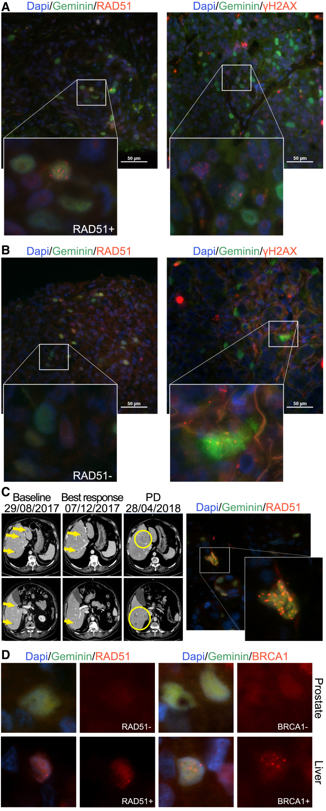
Metastatic prostate cancer (mPC) is enriched for homologous recombination repair (HRR) gene alterations, which have prognostic and predictive value. Routine clinical implementation of next-generation sequencing (NGS) is still limited. We investigated the association between genomic and functional loss of HRR, using NGS and RAD51 immunofluorescence (RAD51-IF) in 219 primary or metastatic biopsies from 187 patients with stage IV prostate cancer. NGS showed frequent genomic alterations in TP53 (40%), AR (15%), PTEN (14%), FOXA1 (12%), MYC (10%), BRCA2 (9%), ATM (8%), and BRCA1 (2%). We pursued RAD51-IF in 206 samples; of those, 139/206 (67%) were evaluable. 21% of samples had RAD51-low score compatible with HRR deficiency (HRD). RAD51-IF showed high sensitivity (71%) and specificity (86%) for BRCA1/2 alterations. Patients with RAD51-low scores experienced longer progression-free survival (PFS) on poly(ADP-ribose) polymerase inhibitors (PARPi) or platinum chemotherapy. RAD51-IF is feasible in routine clinical samples from patients with mPC and is associated with clinically relevant HRR gene alterations.

Keywords: BRCA2; HRR; PARPi; genomics; immunofluorescence; precision medicine; prostate cancer.


Conflict of interest statement

Declaration of interests R.M.-B. reports serving in an advisory role for MSD, Pfizer, Merck, Janssen, and Astellas Pharma and receiving honoraria or travel expenses from Roche, Sanofi Aventis, Astellas, Janssen, MSD, Bayer, Merck, and Pfizer. A.L.-G. and V.S. are co-inventors of a patent related to this work (WO2019122411A1). A.L.-G. is a current employee of AstraZeneca. V.S. has served as an advisor for GSK. J.M. has served as an advisor for AstraZeneca, Amunix/Sanofi, Daichii Sankyo, Janssen, MSD, Pfizer, and Roche; he is a member of the scientific board for Nuage Therapeutics and is involved as an investigator in several pharma-sponsored clinical trials, none of them related to this work.

Figures

None
Graphical abstract
Figure 1
Figure 1
Sample disposition in this study, per assay (A) Diagram of the overall study population and overview of sample disposition. Prim, primary tumor; met, metastatic tumor; HSPC, hormone-sensitive prostate cancer; CRPC, castration-resistant prostate cancer; WES, whole-exome sequencing. (B) Venn diagram for evaluable samples for NGS and/or RAD51-IF assays (WES n = 80, targeted panel n = 139, RAD51-IF n = 139).
Figure 2
Figure 2
Landscape of genomic alterations in the study cohort (n = 181) Oncoprint of pathogenic mutations and copy-number changes (amplifications and homozygous deletions) across the entire cohort grouped by hormone sensitivity status. HSPC, hormone-sensitive prostate cancer; CRPC, castration-resistant prostate cancer. See also Figures S1 and S2.
Figure 3
Figure 3
Distribution of RAD51-IF scores across the study population (A) Bar plot depicting RAD51 foci (bars) and γH2AX (dots) percentage of positive cells across the evaluable samples. (B) Comparison of percentages of γH2AX-positive cells (top) and RAD51-positive cells (lower) in primary vs. metastatic biopsies (left) or based on the hormonal therapy exposure of the patient at the time of biopsy acquisition (hormone naive vs. CRPC, right). Each dot and bar represents one sample. Horizontal orange line indicates the median. ns, no significant difference. See also Table S2.
Figure 4
Figure 4
Association between RAD51-IF results with genomics and clinical outcomes (A) Oncoprint (n = 127) of pathogenic mutations and copy-number changes (amplifications and homozygous deletions), sorted by RAD51 scores from 0 (left) to 100% (right) positive cells in the sample. Homologous recombination repair (HRR) status was determined according to the alteration profile in this pathway, categorizing samples into altered (at least one gene altered, excluding amplifications) or wild type (WT). (B) Kaplan-Meier estimates of progression-free survival (n = 15). In this subset, patients included received PARPi or platinum chemotherapy as monotherapy, had a RAD51-IF-evaluable sample, and the specimen was obtained before treatment start. RAD51 low was defined according to the predefined threshold (≤10%). Progression-free survival is presented as number of months (median, with 95% confidence interval). NE, not estimable. See also Figures S3 and S4–S6; Tables S6 and S7.
Figure 5
Figure 5
Representative cases correlating RAD51-IF score and genomic characterization Images of the RAD51 and yH2AX staining from (A) a RAD51 high sample with no HRR alterations, (B) a RAD51 low case with a BRCA2 pathogenic mutation, (C) a liver biopsy of a patient with a BRCA2 pathogenic mutation after progression to platinum-based chemotherapy that shows high percentage of cells positive for RAD51 foci; contemporaneous ctDNA analysis demonstrated BRCA2 reversion mutations. CT scan (left) of the liver lesions of the patient from baseline, response, and progression to carboplatin. Liver lesions are highlighted in yellow. Representative image of the RAD51 positivity (right) by IF and (D) prostate and liver biopsies of a patient with a monoallelic somatic BRCA1 mutation detected by NGS. The primary prostate tumor shows RAD51-negative cells, but the liver metastasis shows high RAD51 score, in parallel to BRCA1 expression by IF in this liver lesion, but not in the prostate tumor, suggesting restoration of BRCA1 expression in the metastases. Scale bar: 50 μm.

References

    1. Abida W., Armenia J., Gopalan A., Brennan R., Walsh M., Barron D., Danila D., Rathkopf D., Morris M., Slovin S., et al. Prospective Genomic Profiling of Prostate Cancer Across Disease States Reveals Germline and Somatic Alterations That May Affect Clinical Decision Making. JCO Precis. Oncol. 2017;2017:1–16. doi: 10.1200/PO.17.00029. - DOI - PMC - PubMed
    1. Robinson D., Van Allen E.M., Wu Y.-M., Schultz N., Lonigro R.J., Mosquera J.-M., Montgomery B., Taplin M.-E., Pritchard C.C., Attard G., et al. Integrative Clinical Genomics of Advanced Prostate Cancer. Cell. 2015;161:1215–1228. doi: 10.1016/j.cell.2015.05.001. - DOI - PMC - PubMed
    1. Abeshouse A., Ahn J., Akbani R., Ally A., Amin S., Andry C.D., Annala M., Aprikian A., Armenia J., Arora A., et al. The Molecular Taxonomy of Primary Prostate Cancer. Cell. 2015;163:1011–1025. doi: 10.1016/j.cell.2015.10.025. - DOI - PMC - PubMed
    1. Grasso C.S., Wu Y.-M., Robinson D.R., Cao X., Dhanasekaran S.M., Khan A.P., Quist M.J., Jing X., Lonigro R.J., Brenner J.C., et al. The mutational landscape of lethal castration-resistant prostate cancer. Nature. 2012;487:239–243. doi: 10.1038/nature11125. - DOI - PMC - PubMed
    1. Mateo J., Carreira S., Sandhu S., Miranda S., Mossop H., Perez-Lopez R., Nava Rodrigues D., Robinson D., Omlin A., Tunariu N., et al. DNA-Repair Defects and Olaparib in Metastatic Prostate Cancer. N. Engl. J. Med. 2015;373:1697–1708. doi: 10.1056/NEJMoa1506859. - DOI - PMC - PubMed