. 2025 Jun 5;65(6):2301544.
doi: 10.1183/13993003.01544-2023. Print 2025 Jun.

YAP/TAZ are crucial regulators of macrophage-mediated pulmonary inflammation and fibrosis after bleomycin-induced injury

Affiliations

YAP/TAZ are crucial regulators of macrophage-mediated pulmonary inflammation and fibrosis after bleomycin-induced injury

Masum M Mia et al. Eur Respir J. .

Abstract

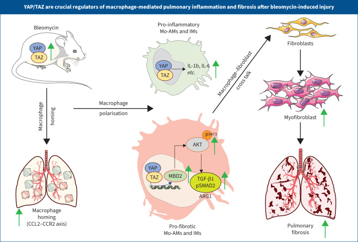
Pulmonary fibrosis is the most prevalent and severe form of end-stage interstitial lung disease. Macrophages are crucial players in inflammation-induced pulmonary fibrosis, but the mechanisms driving macrophage polarisation and their specific roles in pulmonary fibrosis pathogenesis remain poorly understood. Here, we demonstrate that both YAP and TAZ are activated in lung macrophages from patients with pulmonary fibrosis as well as in mice with bleomycin-induced pulmonary fibrosis. Myeloid-specific Yap/Taz deletion resulted in reduced recruitment of monocyte-derived alveolar macrophages (Mo-AMs), impaired inflammatory responses, decreased pulmonary fibrosis and enhanced alveolar epithelial cell regeneration following bleomycin treatment. Conversely, the expression of a constitutively active YAP mutant (YAP5SA) exacerbated bleomycin-induced pulmonary fibrosis by increasing Mo-AM recruitment, elevating expression of pro-inflammatory and pro-fibrotic markers, and impairing alveolar epithelial cell regeneration. We demonstrate that YAP/TAZ-CCL2 (C-C motif chemokine ligand 2) signalling plays a crucial role in bleomycin-induced pulmonary fibrosis, as blocking CCL2 with a neutralising antibody effectively abrogated the YAP5SA-induced recruitment of Mo-AMs, inflammatory and fibrotic responses. Additionally, we reveal that the YAP/TAZ-MBD2-TGFβ1-pSMAD2 signalling axis is crucial not only for pro-fibrotic macrophage polarisation, but also for their cross-talk with lung fibroblasts, driving the fibroblast-to-myofibroblast transition. Collectively, these findings suggest that targeting aberrant YAP/TAZ activity to modulate inflammatory and fibrotic response could be a promising strategy for the prevention and treatment of pulmonary fibrosis.


Conflict of interest statement

Conflict of interest: The authors have no potential conflicts of interest to disclose.

Figures

None
Overview of the study. Mo-AM: monocyte-derived alveolar macrophage; IM: interstitial macrophage; IL: interleukin; CCL: C-C motif chemokine ligand; CCR: C-C motif chemokine receptor; MBD: methyl-CpG–binding domain; TGF: transforming growth factor.
FIGURE 1
FIGURE 1
YAP/TAZ are activated in lung macrophages. a, b) Immunofluorescence staining and quantification of YAP or TAZ co-stained with CD68 on lung sections from human patients diagnosed with pulmonary fibrosis and compared with normal adult human (considered as healthy adult) lung (n=3). c, d) Immunofluorescence staining and quantification of YAP or TAZ co-stained with CD68 in wild-type mouse lung sections at 7 days (D7) post-bleomycin injury compared to saline-treated sham controls (n=5). e, f) Gating strategy and quantification of flow cytometry analysis to sort the total lung macrophages (identified as DapiCD45+Ly6GCD64+CD11b+ cells) using saline-treated sham controls (n=5) and bleomycin-treated mice (n=5) at 7 days. g) Real-time quantitative PCR analysis of Yap, Taz, Ctgf, Il6, Il1β and Tnfα on sorted total lung macrophages. h) Immunoblot analysis of YAP, TAZ and CTGF on primary human lung macrophages treated with saline or lipopolysaccharide (LPS) (100 ng·mL−1) for 12 h (n=3). Quantification of indicated proteins relative to vinculin. i) Immunoblot analysis of YAP and TAZ using cultured primary lung macrophages isolated through flow cytometry (identified as DapiCD45+Ly6GCD64+CD11b+ cells) on saline- or bleomycin-treated control and Yap/Taz double knockout (dKO) mice (n=5) as indicated time points. Quantification of indicated proteins relative to β-actin. SSC: side scatter; FSC: forward scatter; A: area; W: width; H: height; Dapi: 4′,6-diamidino-2-phenylindole; D7: day 7; D14: day 14; Ctgf: connective tissue growth factor; Il: interleukin; TNF: tumour necrosis factor. Data are presented as mean±sem; comparison by two-tailed unpaired t-test. Significance levels are indicated with p-value (significant at p<0.05). *: p<0.05; ***: p<0.001.
FIGURE 2
FIGURE 2
Macrophage-specific Yap/Taz inactivation impairs bleomycin-induced inflammation in mouse lungs. a, b) Immunostaining and quantification of CD68-positive macrophage infiltration in control and double knockout (dKO) lungs treated with saline (day 0 (D0)) or bleomycin for 7 (D7) and 14 days (D14) (n=5). c) ELISA to measure the protein levels of pro-inflammatory cytokines interleukin (IL)-1β, IL-6 and tumour necrosis factor (TNF)-α in bronchoalveolar lavage fluid (BALF) from control and dKO mice (n=5–6) treated with saline (D0) or bleomycin (D7 and D14). d) Real-time quantitative PCR to check the expression of pro-inflammatory cytokines and chemokines including Il6, Il1β, Nos2, Hif1α, Ccl2, Ccr2, Cx3cl1, Cx3cr1, Ccl17, as well as pro-fibrotic markers Arg1 and Fizz1, and the YAP/TAZ target gene Ctgf using lung tissue RNA from control and dKO mice (n=5–8) treated with saline (D0) or bleomycin (D14). Ccl: C-C motif chemokine ligand Nos: nitric oxide synthase; Hif: hypoxia-inducible factor; Ccr: C-C motif chemokine receptor; Cx3cl: C-X3-C motif chemokine ligand; Cx3cr: C-X3-C motif chemokine receptor; Arg: arginase; Fizz: resistin-like molecule-α; Ctgf: connective tissue growth factor; ns: nonsignificant. Data are presented as mean±sem, with statistical comparisons made using a two-tailed unpaired t-test. Significance levels are indicated with p-values (significant at p<0.05).
FIGURE 3
FIGURE 3
Macrophage-specific Yap/Taz inactivation leads to decrease in monocyte-derived alveolar macrophage (Mo-AM) and interstitial macrophage (IM) population after bleomycin-induced injury. a–e) Gating strategy and flow cytometry analysis to sort Mo-AMs (identified as CD45+Ly6GCD64+CD11blowSiglec-Flow), tissue-resident alveolar macrophages (TR-AMs) (identified as CD45+Ly6GCD64+CD11blowSiglec-Fhigh), IMs (CD45+Ly6GCD64+CD11bhighSiglec-F) and neutrophils (identified as CD45+Ly6G+) using lung from control and double knockout (dKO) mice (n=5–9) treated with saline or bleomycin for 7 (D7) and 14 days (D14). f, g) Real-time quantitative PCR for pro-inflammatory cytokines and chemokines such as Il6, Il1β, Ccl2 and Cx3cr1 on sorted Mo-AMs or IMs from control and dKO mice (n=5) treated with saline or bleomycin for 14 days. h, i) Immunoblot analysis of YAP, TAZ, cleaved caspase-1, cleaved interleukin (IL)-1β, nucleotide-binding oligomerisation domain-like receptor containing pyrin domain (NLRP)3, pro-caspase-1, pro-IL-1β and ASC using cell lysates or supernatants as indicated from cultured lung macrophages isolated through flow cytometry (identified as DapiCD45+Ly6GCD64+CD11b+ cells) on bleomycin-treated control and dKO mice (n=5–6). Quantification of indicated proteins relative to β-actin. SSC: side scatter; FSC: forward scatter; A: area; W: width; H: height; Dapi: 4′,6-diamidino-2-phenylindole; Ccl: C-C motif chemokine ligand; Cx3cr: C-X3-C motif chemokine receptor; ns: nonsignificant. Data are presented as mean±sem, with statistical comparisons made using a two-tailed unpaired t-test. Significance levels are indicated with p-value (significant at p<0.05).
FIGURE 4
FIGURE 4
Macrophage-specific Yap/Taz inactivation reduces lung fibrosis after bleomycin-induced injury. a, b) Sirius Red/Fast Green staining and quantification (presented as Ashcroft score) on lung sections from control and double knockout (dKO) mice (n=6–7) treated with saline or bleomycin for 7 (D7) and 14 days (D14), respectively. c) Quantification of hydroxyproline content using lung tissue from control and dKO mice (n=5–7) treated with saline or bleomycin for 14 days. d) Real-time quantitative PCR for pro-fibrotic genes Acta2, Col1a1, Col3a1 and Fn1 on lung tissue from control and dKO mice (n=5–8) treated with saline or bleomycin for 14 days. e, f) Immunostaining and quantification of pro-surfactant protein C (SPC)+alveolar epithelial type-2 (AT2)-cells in control versus dKO mouse lung (n=5) treated with saline or bleomycin for 7 and 14 days. g) Flow cytometry analysis to determine the changes in the number of AT2-cells identified as CD45CD31CD326+MHCII+ in dKO lungs compared to controls (n=5) after saline or bleomycin treatment for 7 and 14 days. h, i) Immunostaining and quantification of Hopx+ AT1-cells in dKO lungs compared to controls (n=5) after saline or bleomycin treatment for 7 and 14 days. Dapi: 4′,6-diamidino-2-phenylindole; MHC: major histocompatibility complex; Acta: smooth muscle actin-α; Col: collagen; Fn: fibronectin; ns: nonsignificant. Data are presented as mean±sem, with statistical comparisons made using a two-tailed unpaired t-test. Significance levels are indicated with p-value (significant at p<0.05).
FIGURE 5
FIGURE 5
Macrophage-specific Yap overexpression augments bleomycin-induced inflammatory response in mice lung. a, b) Immunostaining and quantification of CD68+ macrophage infiltration on the lung from R26YAP5SA and LysMCre;R26YAP5SA mice (n=5) treated with saline or bleomycin for 14 days. c, d) Real-time quantitative (q)PCR for pro-inflammatory cytokines and chemokines such as Il6, Il1β, Hif1α, Arg1, Ccl2, Ccr2, Cx3cl1 and Cx3cr1; with hippo target gene connective tissue growth factor (Ctgf) using lung tissue RNA isolated from R26YAP5SA and LysMCre;R26YAP5SA mice (n=5) treated with saline or bleomycin for 14 days. e–g) Flow cytometry analysis to sort the alveolar macrophages (Mo-AMs) (identified as CD45+Ly6GCD64+CD11blowSiglec-Flow), tissue-resident alveolar macrophages (TR-AMs) (identified as CD45+Ly6GCD64+CD11blowSiglec-Fhigh) and interstitial macrophages (IMs) (CD45+Ly6GCD64+CD11bhighSiglec-F) using lung from R26YAP5SA and LysMCre;R26YAP5SA mice (n=5–6) treated with saline or bleomycin for 14 days. h, i) Real-time qPCR for pro-inflammatory cytokines and chemokines such as Il6, Il1β, Cx3cr1 and Ccl2 on sorted Mo-AMs or IMs from R26YAP5SA and LysMCre;R26YAP5SA mice (n=5) treated with saline or bleomycin for 14 days. Il: interleukin; Hif: hypoxia-inducible factor; Arg: arginase; Ccl: C-C motif chemokine ligand; Ccr: C-C motif chemokine receptor; Cx3cr: C-X3-C motif chemokine receptor; Ctgf: connective tissue growth factor; ns: nonsignificant. Data are presented as mean±sem, with statistical comparisons made using a two-tailed unpaired t-test. Significance levels are indicated with p-value (significant at p<0.05).
FIGURE 6
FIGURE 6
Macrophage-specific Yap overexpression augments bleomycin-induced lung fibrosis in mice. a–c) Sirius Red/Fast Green staining and quantification (presented as Ashcroft score) on lung sections from R26YAP5SA and LysMCre;R26YAP5SA mice (n=5–7) treated with saline or bleomycin for 14 days. d) Quantification of hydroxyproline content using lung tissue from R26YAP5SA and LysMCre;R26YAP5SA mice (n=5–7) treated with saline or bleomycin for 14 days. e) Real-time quantitative (q)PCR for pro-fibrotic genes Acta2, Col1a1, and Col3a1 on lung tissue from R26YAP5SA and LysMCre;R26YAP5SA mice (n=5) treated with saline or bleomycin for 14 days. f–h) Immunoblot analysis and quantification of collagen I and α-smooth muscle actin (SMA) protein using lung tissue from R26YAP5SA and LysMCre;R26YAP5SA mouse (n=5) treated with saline or bleomycin for 14 days. i, j) Immunostaining and quantification of pro-surfactant protein C (SPC)+ alveolar epithelial type-2 (AT2) cells in R26YAP5SA versus LysMCre;R26YAP5SA mice lung (n=5) treated with saline or bleomycin for 14 days. k) Flow cytometry analysis to determine the changes in the number of AT2-cells identified as CD45CD31CD326+MHCII+ in LysMCre;R26YAP5SA lungs compared to R26YAP5SA lungs (n=5) after saline or bleomycin treatment for 14 days. l, m) Immunostaining and quantification of Hopx+ AT1-cells in LysMCre;R26YAP5SA lungs compared to R26YAP5SA lungs (n=5) after saline or bleomycin treatment for 14 days. Acta: smooth muscle actin-α; Col: collagen; Dapi: 4′,6-diamidino-2-phenylindole; MHC: major histocompatibility complex; ns: nonsignificant. Data are presented as mean±sem, with statistical comparisons made using a two-tailed unpaired t-test. Significance levels are indicated with p-values (significant at p<0.05).
FIGURE 7
FIGURE 7
YAP/TAZ modulates C-C motif chemokine ligand (CCL)2 expression and neutralising of CCL2 attenuates YAP-induced pulmonary fibrosis after bleomycin-injury. a) Immunoblot analysis and quantification of CCL2 protein using lung tissue from control and double knockout (dKO) mice (n=5–6) treated with saline or bleomycin for 14 days. b) Immunoblot analysis and quantification of CCL2 protein using lung tissue from R26YAP5SA and LysMCre;R26YAP5SA mice (n=5) treated with saline or bleomycin for 14 days. c) Chromatin immunoprecipitation assay performed on Ccl2 promoter using chromatin from wild-type bone marrow derived macrophages exposed to lipopolysaccharide (100 ng·mL−1) using IgG and YAP or TAZ antibody. d) Graphical presentation of experimental design; R26YAP5SA and LysMCre;R26YAP5SA mice were challenged to intratracheal administration of bleomycin (as 2.5 mg·kg−1 bodyweight (BW)) followed by intraperitoneal injection of CCL2 neutralising antibody or IgG (as 2 mg·kg−1 BW) as indicated time points. e) Immunostaining and quantification of CD68-positive macrophage infiltration in R26YAP5SA and LysMCre;R26YAP5SA mouse lung (n=5) treated with bleomycin followed by IgG or CCL2 neutralising antibody treatment for 14 days. f) Quantification of flow cytometry analysis to sort alveolar macrophages (Mo-AMs), tissue-resident alveolar macrophages (TR-AMs) and interstitial macrophages (IMs) using lung from R26YAP5SA and LysMCre;R26YAP5SA mice (n=7–9) treated with bleomycin followed IgG or CCL2 neutralising antibody treatment for 14 days. g, h) Sirius Red/Fast Green staining and quantification (presented as Ashcroft score) on lung sections from R26YAP5SA and LysMCre;R26YAP5SA mice (n=5) treated with bleomycin following IgG or CCL2 neutralising antibody treatment for 14 days. i) Immunoblot analysis and quantification of collagen III protein in lung tissue from R26YAP5SA and LysMCre;R26YAP5SA mice (n=5) treated with bleomycin followed by IgG or CCL2 neutralising antibody treatment for 14 days. j) mRNA analysis of Il6, Il1β, Acta2, Col1a1 and connective tissue growth factor (Ctgf) on lung tissue from R26YAP5SA and LysMCre;R26YAP5SA mice (n=5) treated with bleomycin followed IgG or CCL2 neutralising antibody treatment for 14 days. k, l) Immunostaining and quantification of Hopx+ AT1-cells in R26YAP5SA and LysMCre;R26YAP5SA lungs (n=5) treated with bleomycin following IgG or CCL2 neutralising antibody treatment for 14 days. m, n) Immunostaining and quantification of pro-surfactant protein C (SPC)+ alveolar epithelial type-2 (AT2) cells in R26YAP5SA and LysMCre;R26YAP5SA mice (n=5) treated with bleomycin followed with IgG or CCL2 neutralising antibody treatment for 14 days. o) Flow cytometry analysis to determine the changes in the number of AT2-cells identified as CD45CD31CD326+ major histocompatibility complex (MHC)II+ in R26YAP5SA and LysMCre;R26YAP5SA lungs (n=7–9) treated with bleomycin followed IgG or CCL2 neutralising antibody treatment for 14 days. D: day; Dapi: 4′,6-diamidino-2-phenylindole; Il: interleukin; Acta: smooth muscle actin-α; Col: collagen; Ctgf: connective tissue growth factor; ns: nonsignificant. Data are presented as mean±sem, with statistical comparisons made using a two-tailed unpaired t-test. Significance levels are indicated with p-value (significant at p<0.05).
FIGURE 8
FIGURE 8
Loss of Yap/Taz attenuates methyl-CpG-binding domain (MBD)2 expression in mouse fibrotic lung and MBD2 acts downstream of YAP during pro-fibrotic macrophage polarisation. a) MBD2 is highly expressed in CD68+ lung macrophages of a patient with pulmonary fibrosis (PF) compared to normal adult human (healthy adult) subject (n=3). b) MBD2 is highly expressed in CD68+ lung macrophages after bleomycin treatment compared to saline treatment in wild-type mice (n=5). c, d) Immunoblot analysis and quantification of MBD2 and arginase (ARG)1 protein using lung tissue from control and double knockout (dKO) mice (n=5) treated with saline or bleomycin for 14 days. e, f) Immunoblot analysis and quantification of MBD2 and ARG1 protein using lung tissue from R26YAP5SA and LysMCre;R26YAP5SA mice (n=5) treated with saline or bleomycin for 14 days. g) Chromatin immunoprecipitation assay performed on Mbd2 promoter using chromatin from wild-type bone marrow-derived macrophages exposed to lipopolysaccharide LPS (100 ng·mL−1) using IgG and YAP or TAZ antibody. h) Real-time quantitative (q)PCR for Mbd2, Arg1 and Tgfβ1 on sorted alveolar macrophages (Mo-AMs) from control and dKO mice (n=5) treated with saline or bleomycin for 14 days. i, j) Immunoblot analysis and quantification of MBD2, ARG1, pAKT (S473), AKT, mature-transforming growth factor (TGF)-β1 and YAP using total lung macrophages (CD45+Ly6GCD64+CD11b+) isolated using flow cytometry and subsequently cultured derived from LysMCre;R26YAP5SA mouse lung (n=5) treated with bleomycin or saline to perform the knockdown experiment with Mbd2 siRNA or control (Ctr) siRNA. k, l) R26YAP5SA and LysMCre;R26YAP5SA mice were challenged to intratracheal administration of bleomycin (as 2.5 mg·kg−1 bodyweight) and the total lung macrophages were harvested and cultured to collect the conditioned medium (CM). k) Immunofluorescence staining and quantification of α-smooth muscle actin (SMA) and collagen I using wild-type mouse lung (n=5) fibroblasts treated with either DMEM or CM collected from R26YAP5SA and LysMCre;R26YAP5SA lung macrophages challenged to bleomycin-injury. l) Immunofluorescence staining and quantification of α-SMA and collagen I using wild-type mouse lung (n=5) fibroblasts treated with IgG or TGFβ1-neutralising antibody (neu ab; 1 μg·mL−1) under the exposure of CM collected from LysMCre;R26YAP5SA lung macrophages challenged to bleomycin-injury. m) Immunofluorescence staining and quantification of α-SMA and collagen I using LysMCre;R26YAP5SA lung (n=5) fibroblasts treated with CM collected from LysMCre;R26YAP5SA lung macrophages challenged to bleomycin-injury followed by Mbd2 siRNA or control siRNA exposure. n) Immunoblot analysis and quantification of pSMAD2 using LysMCre;R26YAP5SA lung fibroblasts treated with either DMEM or CM collected from LysMCre;R26YAP5SA lung macrophages challenged to bleomycin-injury followed by Mbd2 siRNA or control siRNA exposure. The data are represented as mean±sem; comparison by two-tailed unpaired t-test. Significance levels are indicated with p-values (significant at p<0.05). *: p<0.05; ***: p<0.001.
FIGURE 9
FIGURE 9
Graphical model for the macrophage-mediated role of YAP/TAZ during pulmonary fibrosis. Schematic representation showing that development of fibrosis associated with the homing of pro-inflammatory and pro-fibrotic macrophages through the CCL2–CCR2 axis after bleomycin treatment. Pulmonary fibrosis augments the activation of YAP and TAZ in alveolar macrophages (Mo-AMs) and interstitial macrophages (IMs), which induces the release of pro-inflammatory cytokines such as interleukin (IL)-1β, IL-6, etc., as well as pro-fibrotic cytokine transforming growth factor (TGF)-β1 through the MBD2-pAKT-pSMAD2-ARG1 axis that activate the transition of fibroblasts into myofibroblasts leading to pulmonary fibrosis as featured by the expressive production of collagen I, α-smooth muscle actin (SMA) and fibrotic score. Macrophage-specific genetic ablation of Yap/Taz reduces the homing of pro-inflammatory and pro-fibrotic macrophages and defective myofibroblasts formation with impaired pulmonary fibrosis. CCL: C-C motif chemokine ligand; CCR: C-C motif chemokine receptor; MBD: methyl-CpG-binding domain; ARG: Arginase.

Comment in

References

    1. Lederer DJ, Martinez FJ. Idiopathic pulmonary fibrosis. N Engl J Med 2018; 378: 1811–1823. doi:10.1056/NEJMra1705751 - DOI - PubMed
    1. Barratt SL, Creamer A, Hayton C, et al. . Idiopathic pulmonary fibrosis (IPF): an overview. J Clin Med 2018; 7: 201. doi:10.3390/jcm7080201 - DOI - PMC - PubMed
    1. Chen JY, Qiao K, Liu F, et al. . Lung transplantation as therapeutic option in acute respiratory distress syndrome for coronavirus disease 2019-related pulmonary fibrosis. Chin Med J 2020; 133: 1390–1396. doi:10.1097/CM9.0000000000000839 - DOI - PMC - PubMed
    1. Ley B, Collard HR, King TE Jr. Clinical course and prediction of survival in idiopathic pulmonary fibrosis. Am J Respir Crit Care Med 2011; 183: 431–440. doi:10.1164/rccm.201006-0894CI - DOI - PubMed
    1. Hussell T, Bell TJ. Alveolar macrophages: plasticity in a tissue-specific context. Nat Rev Immunol 2014; 14: 81–93. doi:10.1038/nri3600 - DOI - PubMed

MeSH terms

LinkOut - more resources