. 2025 Feb 6;15(1):4535.
doi: 10.1038/s41598-025-88063-8.

Three new species of Mesobiotus (Eutardigrada: Macrobiotidae) from Sweden with an updated phylogeny of the genus

Affiliations

Three new species of Mesobiotus (Eutardigrada: Macrobiotidae) from Sweden with an updated phylogeny of the genus

Sarah Atherton et al. Sci Rep. .

Abstract

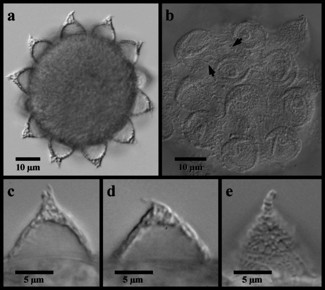
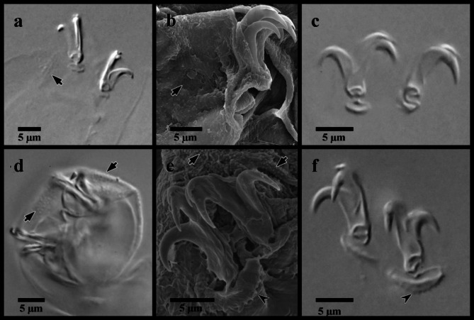
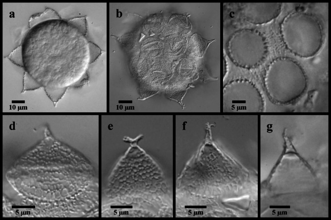
Three new species of Mesobiotus (Tardigrada: Eutardigrada: Macrobiotidae) are described from Skåne County in the southernmost region of Sweden. All three species are distinguished morphologically and through differences in DNA sequences as supported by PTP and mPTP analyses. With the addition of Mesobiotus bockebodicus sp. nov., M. skanensis sp. nov., and M. zelmae sp. nov., the number of nominal species of Macrobiotidae in Sweden has increased to 26, 73% of which have been documented from Skåne. Finally, new morphological details and DNA sequences are presented for Mesobiotus emiliae, a new record is presented of M. mandalori from Sweden, and the phylogenetic relationships within the genus is reconstructed using previously published and new 18S and COI gene sequences.

Keywords: Diversity; Macrobiotidae; New species; Phylogeny; Tardigrades; Taxonomy.


Conflict of interest statement

Declarations. Competing interests: The authors declare no competing interests.

Figures

Fig. 1
Fig. 1
Concatenated 18S and COI gene tree summary with results from the PTP and mPTP analyses. Ultrafast bootstrap support is given at the nodes and species with newly generated genetic data are highlighted in red.
Fig. 2
Fig. 2
Mesobiotus emiliae. (a) whole body, live animal in water; (b) ventral oral cavity armature in Hoyer’s medium; (c) placoid morphology, animal in water; (d) SEM image of the body cuticle; (e) claws and legs IV in Hoyer’s medium, asterisk indicates the larger granulation visible in LM. Black flat arrowhead indicates the constriction of the third macroplacoid; white flat arrowhead indicates examples of the fine granulation present over the entire cuticle.
Fig. 3
Fig. 3
Mesobiotus emiliae eggs. (a) whole egg at the midsection; (b) egg processes and surface; (c,d,e) examples of egg processes; black full arrow indicates crown of thickening at the base of the processes.
Fig. 4
Fig. 4
Mesobiotus mandalori from Sweden (a) whole body, live animal in water; (b) bucco-pharyngeal apparatus, lateral view in Hoyer’s medium; (c) dorsal oral cavity armature in water; (d) ventral oral cavity armature in Hoyer’s medium; (e) placoid morphology, dorsal view in water; (f) placoid morphology, lateral view in water; (g) placoid morphology, dorsal view in Hoyer’s medium. Black full arrows indicate first band of teeth; black flat arrowheads indicate the location of the constriction of the third macroplacoid.
Fig. 5
Fig. 5
Mesobiotus mandalori from Sweden claws. (a) claws of leg II in Hoyer’s medium; (b) SEM image of claws and leg II; (c) claws of leg I in Hoyer’s medium; (d) legs IV in water; (e) SEM image of the claws of leg IV; (f) claws of leg IV in Hoyer’s medium. Black full arrows indicate granulation present on all legs; black indented arrowheads indicate the dentate lunules of the claws of leg IV.
Fig. 6
Fig. 6
Mesobiotus mandalori from Sweden eggs. (a) whole egg at the midsection; (b) surface of the egg and egg processes; (c) bases of the processes and egg interprocess surface; (d,e,f,g) examples of egg processes.
Fig. 7
Fig. 7
Mesobiotus bockebodicus sp. nov. (a,d,f) photo of live holotype specimen 22–046 in water; (b,i) holotype specimen 22–046 in Hoyer’s medium; (c,e) specimen 22-047f in water; (g,h) specimen 24–076 in water. (a) whole body; (b) dorsal oral cavity armature; (c) ventral oral cavity armature; (d) placoid morphology, lateral view; (e) placoid morphology, dorsal view; (f) claws and leg I; (g) claws and leg III; (h,i) claws and legs IV. Black full arrows indicate first band of teeth; black flat arrowheads indicate the location of the constriction of the third macroplacoid; black indented arrowheads indicate the cuticular bars; the white flat arrowhead indicates the cuticular bulge; the asterisk indicates the large granulation present on legs IV.
Fig. 8
Fig. 8
Mesobiotus bockebodicus sp. nov. eggs. (a,d,e) specimen 22–048; (b,f) specimen 22–039; (c) specimen 22–043; (a) whole egg at the midsection; (b) egg surface; (c,d,e,f) examples of egg processes.
Fig. 9
Fig. 9
Mesobiotus skanensis sp. nov. (a,c) holotype specimen 22–163-1, DIC photo of live animal in water; (b,d) specimen 22–159 in water, DIC; (e) paratype specimen 22-118af in Hoyer’s medium, phase contrast. (a) whole body; (b) whole body with a focus on the dorsal body cuticle; (c) ventral body cuticle; (d,e) dorsal body cuticle. The white flat arrowheads indicate examples of the patches of medium-sized granules that appeared present on the body cuticle; the black flat arrowheads indicate examples of individual granules present between the granule patches.
Fig. 10
Fig. 10
Mesobiotus skanensis sp. nov. (a,e,f,) photo of holotype specimen 22–163-1 in Hoyer’s medium, DIC; (b,g) paratype specimen 22–163-5 in Hoyer’s medium, DIC; (c,d) of live holoype specimen 22–163-1 in water, DIC; (h) specimen 22-140f in water, DIC (i) specimen 22-142f in water, DIC. (a) ventral oral cavity armature; (b) dorsal oral cavity armature; (c) placoid morphology, lateral view; (d) placoid morphology, dorsal view; (e) claws of leg I; (f,h) claws of leg II; (g,i) claws of leg IV. Black full arrows indicate first band of teeth; black flat arrowheads indicate the location of the constriction of the third macroplacoid; black indented arrowheads indicate the cuticular bars; the asterisk indicates granulation present on the legs.
Fig. 11
Fig. 11
Mesobiotus skanensis sp. nov. eggs. (a) specimen 22–147-2, whole egg at the midsection; (b) specimen 22–147-4, egg surface; (c) SEM image of whole egg; (d) specimen 22–147-4, egg process; (e) specimen 22–147-1 egg processes; (g,f) SEM images of the egg processes and surface. Black full arrows indicate examples of the “finger-like projections” between the fully connected branches of the processes; black indented arrowheads indicate examples of larger cells that comprise the egg process reticulation; white flat arrowheads indicate examples of small pores present on the egg processes.
Fig. 12
Fig. 12
Mesobiotus zelmae sp. nov. (a) photo of live holotype specimen 22–215h in water; (b) specimen 22–250-15a fixed in Hoyer’s medium; (c,e) holotype specimen 22–215h in Hoyer’s medium; (d) specimen 22–239 in water. (a) whole body; (b) dorsal oral cavity armature; (c) ventral oral cavity armature; (d) placoid morphology, lateral view; (e) placoid morphology, dorsal view; (f) SEM image of the cuticle showing very fine granulation that is present over the entire body. Black full arrows indicate first band of teeth; black flat arrowheads indicate the location of the constriction of the third macroplacoid.
Fig. 13
Fig. 13
Mesobiotus zelmae sp. nov. claws. (a) specimen 22–250-15a, claws of leg I; (b) specimen 23–050, claws of leg II; (c) SEM image of the claws of leg II; (d) specimen 23–050, claws and legs IV; (e) SEM image of the large granules present on legs IV; (f) SEM image of the posterior claw of legs IV (g) SEM image of the claws and legs IV. Black full arrows indicate fine granules present over the entire body cuticle; black indented arrowheads indicate the cuticular bars; the white flat arrowhead indicates the cuticular bulge; asterisks indicate the large granulation present on legs IV.
Fig. 14
Fig. 14
Mesobiotus zelmae sp. nov. eggs. (a,b,f) specimen 22–250-16; (c) specimen 22–449; (d,e) specimen 22–250-15e. (a) whole egg at the midsection; (b) surface of the egg and egg processes; (c,d,e,f) examples of egg processes.

References

    1. Erdmann, W. & Kaczmarek, Ł. Tardigrades in Space Research – Past and Future. Orig Life Evol. Biosph. 47, 545–553. 10.1007/s11084-016-9522-1 (2016). - PMC - PubMed
    1. Guidetti, R., Rizzo, A., Altiero, T. & Rebecchi, L. What can we learn from the toughest animals of the Earth? Water bears (tardigrades) as multicellular model organisms in order to perform scientific preparations for lunar exploration. Planet. Space Sci.74, 97–102. 10.1016/j.pss.2012.05.021 (2012).
    1. Kaczmarek, Ł. et al. Two new species of the genus Milnesium Doyère, 1840 (Tardigrada, Apochela, Milnesiidae) from Madagascar. Zookeys884, 1–22. 10.3897/zookeys.884.29469 (2019). - PMC - PubMed
    1. Bartylak, T. et al. Terrestrial Tardigrada (Water Bears) of the Słowiński National Park (Northern Poland). Diversity16, 239. 10.3390/d16040239 (2024).
    1. Dueñas-Cedillo, A. et al. Towards an inventory of Mexican tardigrades (Tardigrada): a survey on the diversity of moss tardigrades with an emphasis in conifer forests from the Valley of Mexico Basin. Check List.20, 471–498. 10.15560/20.2.471 (2024).

LinkOut - more resources