. 2025 Feb 6;15(1):4523.
doi: 10.1038/s41598-025-88918-0.

Increasing the concentration of plasma molecules improves the biological activity of platelet-rich plasma for tissue regeneration

Affiliations

Increasing the concentration of plasma molecules improves the biological activity of platelet-rich plasma for tissue regeneration

Mikel Sánchez et al. Sci Rep. .

Abstract

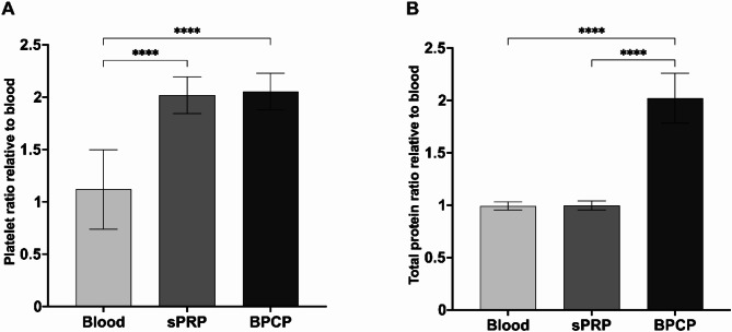
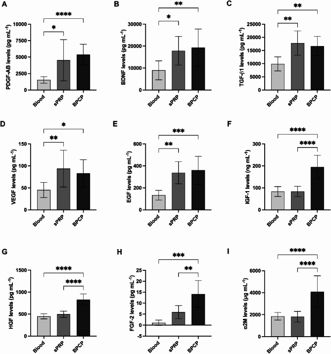
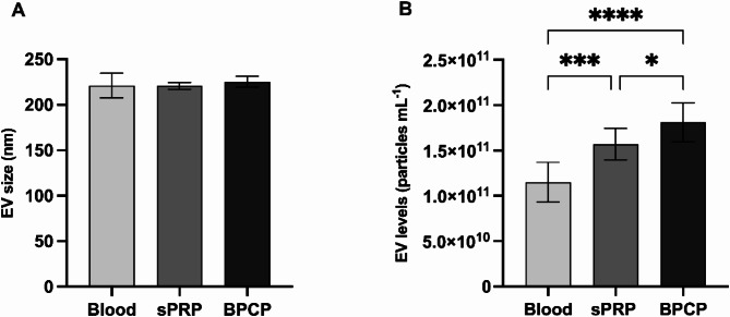
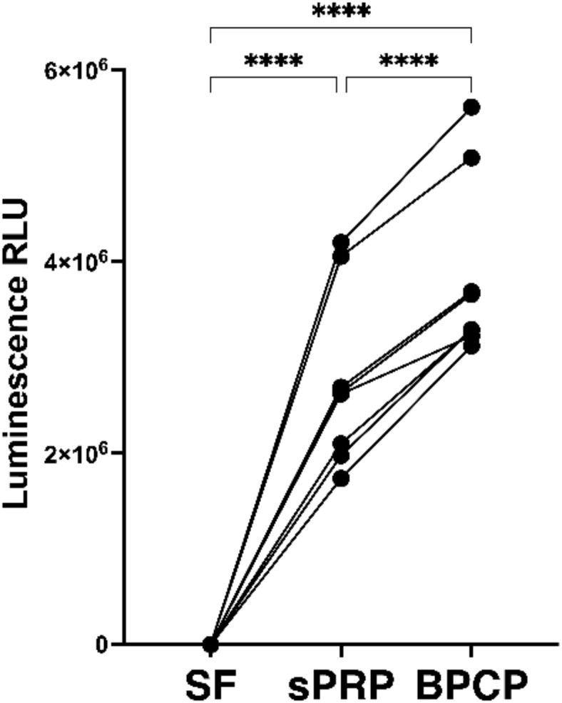
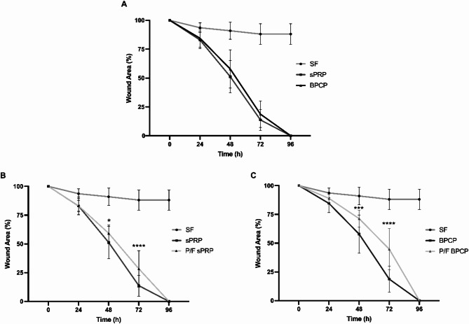
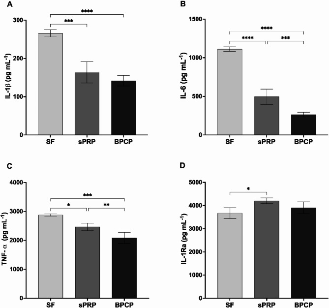
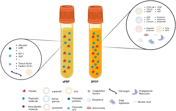
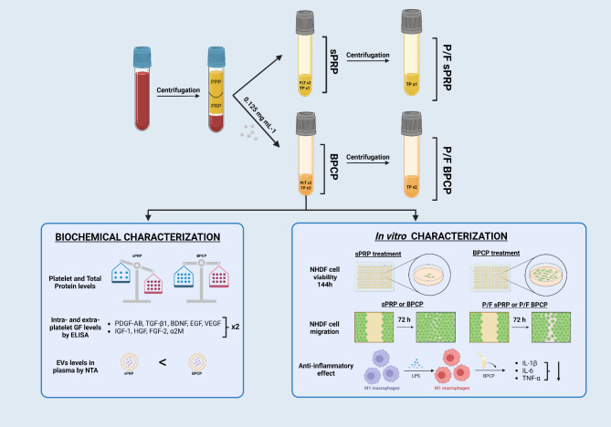
Platelet-rich plasma (PRP) has emerged as a promising therapy in a variety of medical fields. However, it is crucial to go beyond simple platelet concentration and examine the complex molecular composition both inside and outside platelets. The present work studies the effectiveness of a novel type of PRP named 'balanced protein-concentrate plasma' (BPCP). Different growth factor (GF) levels were measured using Enzyme Linked Immunosorbent Assay (ELISA), and in addition to the increase in intra-platelet GFs found in standard PRP (sPRP), BPCP also showed a higher concentration of plasmatic protein. Furthermore, extracellular vesicle (EV) concentration was significantly higher in BPCP. Cell proliferation was higher in cells incubated with lysates derived from BPCP compared to those cultured with sPRP. Regarding cell migration capacity, it was found that the process is platelet-dependent. Finally, the anti-inflammatory effect of BPCP was evaluated by inducing an inflammatory environment in M1-type macrophages. Cytokine levels were measured by ELISA following BPCP administration, showing a significant decrease in pro-inflammatory IL-1β, IL-6 and TNF-α. In summary, although further preclinical and clinical studies are needed in order to determine the therapeutic potential of BPCP, this PRP with unique characteristics demonstrates encouraging in vitro results that could potentially enhance tissue regeneration capacity.

Keywords: Anti-inflammatory effect; Biomolecules; Cell migration; Cell proliferation; Growth factors; Platelet-rich-plasma.


Conflict of interest statement

Declarations. Competing interests: The authors declare the following financial interests/personal relationships which may be considered as potential competing interests: MS has a patent pending with the title “PLASMA ENRICHED IN PLATELETS AND PLASMA MOLECULES” and reference code “PCT/EP2024/059994”. If there are other authors, they declare that they have no known competing financial interests or personal relationships that could have appeared to influence the work reported in this paper.

Figures

Fig. 1
Fig. 1
Platelet and total protein concentration levels in sPRP and BPCP. Mean values of the amount of platelets (A) and total protein (B) in whole blood, sPRP and BPCP are shown. Error bars = standard deviation (n = 10). Statistically significant differences were calculated using one-way ANOVA test (**** p < 0.0001).
Fig. 2
Fig. 2
α2M protein, platelet and extraplatelet GF levels measured by ELISA. Mean values of platelet GFs PDGF-AB (A), BDNF (B), TGF-β1 (C), VEGF (D), EGF (E), the extraplatelet growth factor IGF-1 (F), the intra- and extraplatelet growth factor HGF (G), FGF-2 (H) and the plasmatic protein α2M (I) in blood, sPRP and BPCP are shown. Error bars = standard deviation (n = 6–11). Statistically significant differences were calculated using one-way ANOVA and Kruskal-Walli’s test (* p < 0.05; ** p < 0.01; *** p < 0.001; **** p < 0.0001).
Fig. 3
Fig. 3
Characterisation of platelet-derived EVs by NTA. Particle size (A) and concentration (B) were analysed in blood, sPRP and BPCP. Error bars = standard deviation (n = 9). Statistically significant differences were calculated using one-way ANOVA test (* p < 0.05; *** p < 0.001; **** p < 0.0001).
Fig. 4
Fig. 4
Cellular viability in NHDF cells. The viability levels of NHDF cells incubated with sPRP and BPCP are expressed as relative light units (RLUs), and each line represents a different donor (n = 8). Serum-free (SF) condition served as negative control. Statistical analysis was calculated using one-way ANOVA (p < 0.0001).
Fig. 5
Fig. 5
Wound healing percentage in NHDF cultured with BPCP, sPRP, P/F sPRP and P/F BPCP lysates. Comparisons of percentage of wound width are shown between sPRP and BPCP (A), sPRP and P/F sPRP (B), and BPCP and P/F BPCP (C). Four conditions were run in parallel with SF as a negative control. Error bars = standard deviation (n = 15). Statistically significant differences were calculated using two-way ANOVA test (* p < 0.05; *** p < 0.001; **** p < 0.0001).
Fig. 6
Fig. 6
Analysis of pro- and anti-inflammatory cytokine levels in LPS stimulated M1-type macrophages by ELISA. IL-1β (A), IL-6 (B), TNF-α (C) and IL-1Ra (D) were measured in culture medium of M1-type macrophages treated with sPRP and BPCP under LPS induced inflammatory environment. SF was used as a negative control. Error bars = standard deviation (n = 6). Statistically significant differences were calculated using one-way ANOVA test and Kruskal-Walli’s test (* p < 0.05; ** p < 0.01; *** p < 0.001).
Fig. 7
Fig. 7
Schematical representation of biochemical content of a standard PRP (sPRP) and balanced protein-concentrate plasma (BPCP). The platelet amount represents a two-fold increase in both formulations compared to blood basal levels. Platelets are composed of α-granules, which are rich in growth factors (GFs); dense granules with several hormones, ions and neurotransmitters; and lysosomes, which contain degrading enzymes. Moreover, numerous extracellular vesicles (EVs) are found, such as exosomes (Exos) and microvesicles (MVs), in addition to several organelles. Platelet-derived molecules are represented in red. Nevertheless, plasmatic molecules are doubled in BPCP compared to blood and sPRP. These biomolecules include plasmatic proteins (e.g. albumin, fibrinogen and α2M), GFs (IGF-1 and HGF, for instance) and coagulation factors, among others. Platelet-derived EVs are also found. (Image created with Biorender.com)
Fig. 8
Fig. 8
Diagram of the biochemical and in vitro characterisation of BPCP. BPCP formulation and its respective P/F formulation were obtained. In this study, biochemical characterisation was performed via the analysis of platelets and total protein levels, GF and α2M concentration and EV content. Moreover, in vitro characterisation was performed to assess cell viability, migration and anti-inflammatory capacity. Image created with Biorender.com.
Fig. 9
Fig. 9
NHDF in vitro migration through the artificially made scratch. The pictures were taken immediately after the addition of medium supplemented with sPRP and BPCP (A) and after 48 h (B) under 5x magnification. Wound area (µm2) is represented by the cell-free area.

References

    1. Machado, E. S. et al. Turn down-turn up: Asimple and low-cost protocol for preparing platelet-rich plasma. Clin. (Sao Paulo). 74, e1132 (2019). - PMC - PubMed
    1. Sánchez, M. et al. Isolation, Activation, and Mechanism of Action of Platelet-Rich Plasma and Its Applications for Joint Repair. Regenerative MedicineIntechOpen, (2019). 10.5772/intechopen.90543
    1. Chen, Y., Zhong, H., Zhao, Y., Luo, X. & Gao, W. Role of platelet biomarkers in inflammatory response. Biomark. Res.8, 28 (2020). - PMC - PubMed
    1. van der Meijden, P. E. J. & Heemskerk, J. W. M. Platelet biology and functions: New concepts and clinical perspectives. Nat. Rev. Cardiol.16, 166–179 (2019). - PubMed
    1. Graca, F. A. et al. Platelet-derived chemokines promote skeletal muscle regeneration by guiding neutrophil recruitment to injured muscles. Nat. Commun.14, 2900 (2023). - PMC - PubMed

Substances

LinkOut - more resources