. 2025 Feb;31(2):e70257.
doi: 10.1111/cns.70257.

Caloric Restriction Preserves BBB Integrity After Transient Focal Cerebral Ischemia Through Reducing Neutrophil Infiltration

Affiliations

Caloric Restriction Preserves BBB Integrity After Transient Focal Cerebral Ischemia Through Reducing Neutrophil Infiltration

Chenran Wang et al. CNS Neurosci Ther. 2025 Feb.

Abstract

Aims: Caloric restriction is a health-promoting lifestyle that has been reported to protect both white and gray matter in cases of ischemic stroke. This study will explore the underlying mechanism of restricted feeding (RF) and provide a theoretical basis for precise clinical treatment of stroke.

Methods: In this study, we pretreated C57BL/6J mice with 70% RF for a continuous 28-day period prior to 60 min of transient focal cerebral ischemia (tFCI). Histological staining, diffusion tensor imaging (DTI), and behavioral assessments were used to assess RF's neuroprotection following tFCI. Immunofluorescence staining, quantitative real-time PCR, and flow cytometry were conducted to evaluate brain inflammation post-tFCI. Western blot, immunofluorescence staining, tracers, and electric microscopy were used to assess the blood-brain barrier (BBB) integrity. Peripheral neutrophils were cleared by administrating an anti-Ly6G antibody.

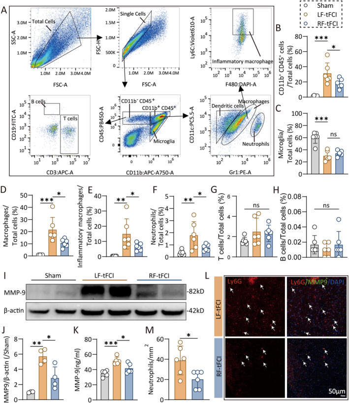
Results: Initially, DTI, NeuN staining, and a batch of behavioral tests verified that RF significantly mitigated both gray/white matter injury and neurological deficits in the short- and long-term following tFCI. RF mice showed more anti-inflammatory microglia in their brains, along with reduced inflammatory cytokines, and chemokines. Interestingly, RF significantly reduced the neutrophils and macrophage infiltration. Subsequently, we observed that RF mice exhibited better BBB integrity following tFCI, with reduced neutrophil infiltration and matrix metalloprotein-9 release. Furthermore, the clearance of neutrophils with anti-Ly6G antibody in ad libitum feeding mice (LF-Ly6G) elicited comparable neuroprotective effects to those observed in RF, including improvements in neurological deficits, reductions in infarct volume, and mitigation of BBB damage.

Conclusions: In summary, our findings suggest that RF maintains the BBB integrity following ischemic stroke at least partially by reducing neutrophil infiltration, thereby alleviating both neurological and histological impairments.

Keywords: blood–brain barrier; caloric restriction; neutrophils; stroke.


Conflict of interest statement

The authors declare no conflicts of interest.

Figures

FIGURE 1
FIGURE 1
Caloric restriction ameliorates histological and neurological deficits following ischemic stroke. (A) Representative images of MAP2 immunofluorescence staining. (B, C) The total atrophy volume (B) and the atrophy area (C) of each layer from rostral to caudal at 28 days after tFCI. n = 6/group. (D) DEC map presented was used to visualize in vivo DTI on Day 14 after tFCI. Scale bar: 3 mm. (E, F) Statistical plots illustrate the variations in FA and RD within the EC (E) and IC (F) regions on Days 3 and 14 post‐tFCI. n = 5/group. (G–I) Sensorimotor deficits were evaluated before (pre) and up to 35 days after tFCI or Sham surgery by Garcia score (G) and grid‐walking test (H, I). n = 6/group. (J–L) Morris water maze test showed the tracking plot of (J), swimming speed (K), and the percentage of distance traveled in target quadrant (L) in test. n = 8–10/group. All data are presented as means ± SEM. Data were analyzed using unpaired two‐tail Student's t‐test (B, C), one‐way (K, L) or two‐way (E, F, G–I) ANOVA followed by Bonferroni post hoc test, and Kruskal–Wallis test with Dunn's multiple‐comparison post hoc test (L). *p < 0.05, **p < 0.01, ***p < 0.001, ns, no significance, as indicated.
FIGURE 2
FIGURE 2
Caloric restriction modulates microglia heterogeneity, and the expression of inflammatory cytokines, and chemokines in short term after ischemic stroke. (A) Schematic diagram indicating the regions where images were captured in (C). (B) Representative images of triple immunostaining (Iba1, CD16/32, and Arg1) taken from the dashed box in Figure 2C. The green arrow indicated promicroglia, red arrow indicated antimicroglia, yellow arrow indicated transit microglia, and purple arrow indicated rest microglia. Scale bar: 20 um. (C) Representative images of Iba1, CD16/32, and Arg1 immunofluorescence staining in striatum 3 days after tFCI. Scale bar: 50 um. (D) The density of Iba1+ cells. n = 6/group. (E‐F) The percentage of different microglia phenotypes accounted for Iba1+ cells. n = 6/group. (G‐H) The relative expression of proinflammatory cytokines (G) and chemokines (H). n = 5/group. All data are presented as means ± SEM. Data were analyzed using unpaired two‐tail Student's t‐test (D–F), one‐way ANOVA followed by Bonferroni post hoc test (H), and Kruskal–Wallis test with Dunn's multiple‐comparison post hoc test (G). *p < 0.05, **p < 0.01, and ***p < 0.001, as indicated.
FIGURE 3
FIGURE 3
Caloric restriction preconditioning reduces peripheral neutrophils and macrophages infiltration in acute phase after ischemic stroke. (A) Gating strategy of flow cytometry that analyzes the infiltrated immune cells in brain 3 days after tFCI. (B–H) The percentage of CD11b+CD45+ cells (B), microglia (C), macrophages (D), inflammatory macrophages (E), neutrophils (F), T cells (G), and B cells (H) accounted for total cells. n = 6/group. (I) Western blot of the penumbra and core tissue 3 days after tFCI were probed with anti‐MMP9 and β‐actin. (J) Quantification of relative MMP9 protein expression in western blot. n = 4/group. (K) ELISA analysis of MMP9 in brain 3 days after tFCI. n = 5/group. (L‐M) Representative images (L) and quantification (M) of Ly6G/MMP9/DAPI immunofluorescence staining 3 days after tFCI. Scale bar, 50 um. n = 6/group. All data are presented as means ± SEM. Data were analyzed using one‐way ANOVA followed by Bonferroni post hoc test (C, G, J, K), Kruskal–Wallis test with Dunn's multiple‐comparison post hoc test (B, D–F, H), and Mann–Whitney test (M). *p < 0.05, **p < 0.01, and ***p < 0.001; ns, no significance, as indicated.
FIGURE 4
FIGURE 4
Caloric restriction protects the blood–brain barrier integrity after ischemic stroke. (A) Images of EB extravasation 48 h after tFCI. (B, C) The content of EB dye in each layer from rostral to caudal (B) and in the whole brain (C). n = 5/group. (D) Representative images of double immunostaining of cadaverine and lectin in cortex and striatum 48 h after tFCI. (E) Quantification of fluorescence intensity of cadaverine. n = 3–4/group. (F) The immunofluorescence images and 3D reconstruction images of IgG 48 h after tFCI. (G, H) Quantification of the leaked area (G) and fluorescence intensity (H) of IgG. n = 5/group. (I–K) The western blot images (I) and the relative expression (J, K) of ZO‐1 and cadherin 3 days after tFCI. n = 3–4/group. (L) Schematic diagram indicating the regions where images were captured in (M). (M) Representative images of CD31/ZO‐1 immunofluorescence 3 days after tFCI. (N) Quantification of fluorescence intensity of ZO‐1. n = 5 (RF‐tFCI group) or 8 (LF‐tFCI group). (O) Electric microscopy images of blood–brain barrier 48 h after tFCI. L: Capillary lumen; TJ: tight junction; *Astrocyte end‐feet; arrow: basement membrane. All data are presented as means ± SEM. Data were analyzed using unpaired two‐tail Student's t‐test (E, N), one‐way (J, K) or two‐way (B) ANOVA followed by Bonferroni post hoc test, or Kruskal–Wallis test with Dunn's multiple‐comparison post hoc test (C, G, H). *p < 0.05, **p < 0.01, ***p < 0.001, # p < 0.05, ## p < 0.01, and ### p < 0.001, as indicated.
FIGURE 5
FIGURE 5
Reducing neutrophil infiltration may contribute to the protective effects of caloric restriction in ischemic stroke. (A) Diagram illustrating the spatiotemporal strategy for Ly6G injection and the following functional and histological measurements. (B) Weight change (vs. weight before surgery, %) after tFCI. Sensorimotor deficits were evaluated before (pre) and up to 7 days after tFCI by Garcia score (C), hang‐wire score (D), and foot faults test (E). n = 5/group. (F, G) Representative images (F) and the quantification of the infarct area (G) 7 days after tFCI. n = 5/group. (H, I) Representative images (H) and the mean immunofluorescent intensity (I) of ZO‐1 in the infarct core. (J‐K) Western blot of the penumbra and core tissue 3 days after tFCI were probed with anti‐MMP9 and β‐actin. n = 4–5/group. All data are presented as means ± SEM. Data were analyzed using one‐way (G, K) or two‐way (B–E) ANOVA followed by Bonferroni post hoc test, or Kruskal–Wallis test with Dunn's multiple‐comparison post hoc test (I). *p < 0.05, **p < 0.01, and ***p < 0.001, ns, no significance, as indicated.

Similar articles

References

    1. “World Stroke Day 2022,” accessed September 2, 2024, https://www.who.int/srilanka/news/detail/29‐10‐2022‐world‐stroke‐day‐2022.
    1. Hilkens N. A., Casolla B., Leung T. W., and de Leeuw F. E., “Stroke,” Lancet 403, no. 10446 (2024): 2820–2836, 10.1016/S0140-6736(24)00642-1. - DOI - PubMed
    1. Campbell B. C. V., De Silva D. A., Macleod M. R., et al., “Ischaemic Stroke,” Nature Reviews Disease Primers 5, no. 1 (2019): 70, 10.1038/s41572-019-0118-8. - DOI - PubMed
    1. Bushnell C., Kernan W. N., Sharrief A. Z., et al., “2024 Guideline for the Primary Prevention of Stroke: A Guideline From the American Heart Association/American Stroke Association,” Stroke; a Journal of Cerebral Circulation 55, no. 12 (2024): e344–e424, 10.1161/STR.0000000000000475. - DOI - PubMed
    1. Zhang M., Che R., Xu J., et al., “Long Term for Patients With Futile Endovascular Reperfusion After Stroke,” CNS Neuroscience & Therapeutics 30, no. 3 (2024): e14588, 10.1111/cns.14588. - DOI - PMC - PubMed

MeSH terms