. 2024 Dec 27:21:76.
doi: 10.25259/Cytojournal_153_2024. eCollection 2024.

Influence of sodium ferulate on neutrophil extracellular traps-platelet activation-mediated endothelial dysfunction in immune small vasculitis

Affiliations

Influence of sodium ferulate on neutrophil extracellular traps-platelet activation-mediated endothelial dysfunction in immune small vasculitis

Xiaoli Zhou et al. Cytojournal. .

Abstract

Objective: Anti-neutrophil cytoplasmic antibody (ANCA)-associated vasculitis (AAV) is an autoimmune disease that is challenging to treat. This study aimed to identify the effect of sodium ferulate on endothelial dysfunction mediated by neutrophil extracellular trap (NET)-platelet activation in AAV to provide potential strategies for AAV treatment.

Material and methods: An animal model of myeloperoxidase (MPO)-AAV passive immune vasculitis was established using anti-MPO immunoglobulin G and Rag2 knockout mice. The efficacy and mechanism of action of sodium ferulate in AAV were explored in cultured and isolated endothelial progenitor cells (EPCs), and messenger ribonucleic acid gene expression, relative protein expression, and protein fluorescence intensity were determined through quantitative polymerase chain reaction, Western blotting, and immunofluorescence, respectively. Serum antibody concentrations were determined by enzyme-linked immunosorbent assay, and flow cytometry was used in determining the expression levels of platelet-selectin (CD62p) and procaspase-activating compound-1 (PAC-1) on the surfaces of the platelets. The EPCs' ultramicroscopic structure was observed through transmission electron microscopy.

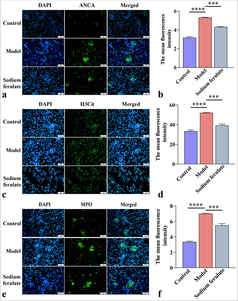
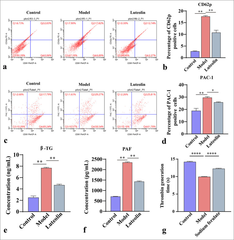
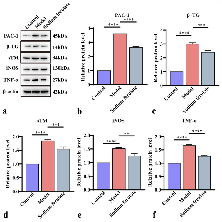
Results: The expression levels of ANCA, histone H3 citrullinated, and MPO protein fluorescence intensity in MPO-AAV mice were inhibited by sodium ferulate, and the expression levels of CD62p and PAC-1 on the cell surface were reduced. The relative expression levels of β-trace protein (β-TG), soluble thrombomodulin, inducible nitric oxide synthase (iNOS), and tumor necrosis factor α decreased. We found that sodium ferulate inhibited NETs' free DNA and mitigated damage in EPCs. In addition, relative expression levels of von Willebrand Factor, β-TG, and iNOS and serum concentrations of PAC-1, β-TG, and iNOS were inhibited.

Conclusion: Sodium ferulate can treat AAV by inhibiting NET release and platelet activation and reducing endothelial cell damage.

Keywords: antineutrophil cytoplasmic antibody; neutrophil extracellular trap; platelet; sodium ferulate; vasculitis.


Conflict of interest statement

The authors declare no conflict of interest.

Figures

Figure 1:
Figure 1:
Immunofluorescence microscopy of ANCA, H3Cit, and MPO. (a and b) ANCA immunofluorescence microscopy observation and its average fluorescence intensity; (c and d) H3Cit immunofluorescence microscopy observation and its average fluorescence intensity; (e and f) MPO immunofluorescence microscopy observation and its average fluorescence intensity at original magnification (×400). Scale bar = 50 μm. Data of three independent experiments are presented as mean ± SD and subjected to ordinary one-way ANOVA (“” indicates statistically significant difference at P < 0.001, and “” indicates extremely significant difference at P < 0.0001. DAPI: 4',6-Diamidino-2-phenylindole, H3Cit: Histone H3 citrullinated, ANCA: Antineutrophil cytoplasmic antibody, MPO: Myeloperoxidas e, ANOVA: Analysis of variance).
Figure 2:
Figure 2:
Platelet activation assessment. (a and b) CD62p, a biomarker of platelet activation, and its percentage of positive cells by flow cytometry; (c and d) PAC-1 B, a biomarker of platelet activation, and its percentage of positive cells by flow cytometry; (e and f) Enzyme-linked immunosorbent assay for β-TG and PAF concentrations in mouse serum; (g) Platelet thrombin generation time was measured by thrombin generation assay. Data of three independent experiments are presented as mean ± SD and subjected to ordinary one-way ANOVA (“” indicates a statistically significant difference at P < 0.05, “” indicates a statistically significant difference at P < 0.01, and “” indicates extremely significant difference at P < 0.0001. CD62P: Platelet-selectin, β-TG: β-Trace protein, PAC-1: Procaspase-activating compound-1, PAF: Platelet-activating factor, ANOVA: Analysis of variance, SD: Standard deviation).
Figure 3:
Figure 3:
Efficacy of sodium ferulate in affecting PAC-1, β-TG, sTM, iNOS, and TNF-α content in the kidney tissues of MPO-AAV mice. (a) Relative mRNA gene level of PAC-1; (b) relative mRNA gene level of β-TG; (c) relative mRNA gene level of sTM; (d) relative mRNA gene level of iNOS; (e) relative mRNA gene level of TNF-α. Data of three independent experiments are presented as mean ± SD and subjected to ordinary one-way ANOVA (“” indicates a statistically significant difference at P < 0.05, “” indicates a statistically significant difference at P < 0.01, “” indicates statistically significant difference at P < 0.001 and “” indicates extremely significant difference at P < 0.0001. PAC-1: Procaspase-activating compound-1, β-TG: β-Trace protein, sTM: Soluble thrombomodulin, iNOS: Inducible nitric oxide synthase, TNF-α: Tumor necrosis factor α, MPO: Myeloperoxidase, AAV: Associated vasculitis, mRNA: Messenger ribonucleic acid, ANOVA: Analysis of variance, SD: Standard deviation).
Figure 4:
Figure 4:
Efficacy of sodium ferulate in affecting content of PAC-1, β-TG, sTM, iNOS, and TNF-α in the kidney tissues of MPO-AAV mice. (a) Western blot detection of the protein bands of PAC-1, β-TG, sTM, iNOS, and TNF-α; (b) grayscale analysis of PAC-1 bands; (c) grayscale analysis of β-TG bands; (d) grayscale analysis of sTM bands; (e) grayscale analysis of iNOS bands; (f) grayscale analysis of TNF-α bands. Data of three independent experiments are presented as mean ± SD and subjected to ordinary one-way ANOVA (“” indicates a statistically significant difference at P < 0.01, “” indicates a highly significant difference at P < 0.001, and “” indicates extremely significant difference at P < 0.0001. PAC-1: Procaspase-activating compound-1, β-TG: β-Trace protein, sTM: Soluble thrombomodulin, iNOS: Inducible nitric oxide synthase, TNF-α: Tumor necrosis factor-α, MPO: Myeloperoxidase, AAV: Associated vasculitis, ANOVA: Analysis of variance, SD: Standard deviation).
Figure 5:
Figure 5:
Effect of sodium ferulate on serum stimulated EPCs. (a) Detection of NETs by fluorescence staining; (b) MTT assay to assess the viability of EPCs; (c) ultrastructure of serum-stimulated EPCs by transmission electron microscopy, where “L-sodium ferulate” denotes low-dose sodium ferulate, “M-sodium ferulate” denotes medium-dose sodium ferulate, and “H-sodium ferulate” denotes high-dose sodium ferulate. MTT: 3-(4,5-Dimethylthiazol-2-yl)-2,5-diphenyltetrazolium bromide assay, EPCs: Endothelial progenitor cells, NETs: neutrophil extracellular traps. Scale bar = 1 μm. Data of three independent experiments are presented as mean ± SD and subjected to ordinary one-way ANOVA (Significance was represented by asterisks: , P < 0.05, , P < 0.001, , P < 0.0001 versus control. ####, P < 0.0001 versus model, ANOVA: Analysis of variance, SD: Standard deviation).
Figure 6:
Figure 6:
Effect of sodium ferulate on serum stimulated EPCs. (a) Concentration of PAC-1 in the supernatant of stimulated EPCs; (b) concentration of β-TG in the supernatant of stimulated EPCs; (c) concentration of iNOS in the supernatant of stimulated EPCs; (d) Western blotting to detect the protein bands of vWF, β-TG, and iNOS; (e) analysis of the gray values of the protein bands of vWF, β-TG, and iNOS. Relative analysis was made based on the gray value of the strip chart; β-actin employed as the endogenous control, where “L-sodium ferulate” denotes low-dose sodium ferulate, “M-sodium ferulate” denotes medium-dose sodium ferulate, and “H-sodium ferulate” denotes high-dose sodium ferulate. Data of three independent experiments are presented as mean ± SD and subjected to ordinary one-way ANOVA.Significance is indicated by asterisks: , P < 0.05, , P < 0.01; , P < 0.001, , P < 0.0001 compared with the control. “ns” indicates no statistical significance compared with the model, ##, P < 0.01 ###, P < 0.001, ####, P < 0.0001 versus model (PAC-1: Procaspase-activating compound-1, β-TG: β-Trace protein, iNOS: Inducible nitric oxide synthase, vWF: von Willebrand Factor, ANOVA: Analysis of variance, EPCs: Endothelial progenitor cells, SD: Standard deviation).

References

    1. Nakazawa D, Masuda S, Tomaru U, Ishizu A. Pathogenesis and therapeutic interventions for ANCA-associated vasculitis. Nat Rev Rheumatol. 2019;15:91–101. doi: 10.1038/s41584-019-0168-z. - DOI - PubMed
    1. Kitching AR, Anders HJ, Basu N, Brouwer E, Gordon J, Jayne DR, et al. ANCA-associated vasculitis. Nat Rev Dis Primers. 2020;6:71. doi: 10.1038/s41572-020-0204-y. - DOI - PubMed
    1. Guchelaar NA, Waling MM, Adhin AA, van Daele PL, Schreurs MW, Rombach SM. The value of anti-neutrophil cytoplasmic antibodies (ANCA) testing for the diagnosis of ANCA-associated vasculitis, a systematic review and meta-analysis. Autoimmun Rev. 2021;20:102716. doi: 10.1016/j.autrev.2020.102716. - DOI - PubMed
    1. Ríos-López AL, González GM, Hernández-Bello R, SánchezGonzález A. Avoiding the trap: Mechanisms developed by pathogens to escape neutrophil extracellular traps. Microbiol Res. 2021;243:126644. doi: 10.1016/j.micres.2020.126644. - DOI - PubMed
    1. Liu L, Mao Y, Xu B, Zhang X, Fang C, Ma Y, et al. Induction of neutrophil extracellular traps during tissue injury: Involvement of STING and Toll-like receptor 9 pathways. Cell Prolif. 2019;52:e12579. doi: 10.1111/cpr.12579. - DOI - PMC - PubMed

LinkOut - more resources