

A new technology for a novel clinical approach in a dog with a complex vascular anomaly: the "extended reality"
- PMID: 39918737
- PMCID: PMC11805765
- DOI: 10.1007/s11259-025-10668-1
A new technology for a novel clinical approach in a dog with a complex vascular anomaly: the "extended reality"
Abstract
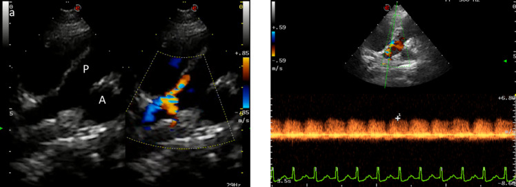
Extended reality includes both virtual and augmented realities. In virtual reality objects are rendered in an artificial environment where the user can move and interact with a head mounted display. In augmented reality virtual objects are superimposed to real environment enriching it via a head mounted display. In human medicine these technologies have been already used for educational surgical purposes, but remain relatively unknown in veterinary medicine. We report a case of a 1-year-old, female, French bulldog presented for exercise intolerance and dyspnea. Echocardiography showed signs of left ventricular enlargement with reduced fractional shortening and turbulent flow distal to the pulmonary artery bifurcation. Computed tomography revealed a complex vascular network comprising the descending aorta and left pulmonary artery resembling a patent ductus arteriosus. Virtual reality was used for the surgical planning and a left thoracotomy was performed to close the abnormal vessel at the level of the entrance in the left pulmonary artery with augmented reality assistance. No complications were reported during or after the surgery and the dog completely recovered. Echocardiographic findings 3 days, 1 month and 18 months after the surgery demonstrated absence of residual flow and improving ventricular dimensions. To our knowledge this report documents the first use of extended reality for the visualization, planning and execution of the surgical correction of a complex vascular defect in veterinary medicine.
Keywords: Augmented reality; Canine; Cardiology; Vascular surgery; Virtual reality.
© 2025. The Author(s).
Conflict of interest statement
Declarations. Ethics approval: Ethical review and approval were waived for this study because, being a case report derived from clinical practice, all medical procedures were performed after obtaining written consent from the owner. Competing interests: The authors declare no competing interests.
Figures


References
-
- Adballah M, Espinel Y, Calvet L et al (2022) Augmented reality in laparoscopic liver resection evaluated on an ex-vivo animal model with pseudo-tumours. Surg Endosc 36:833–843. 10.1007/s00464-021-08798-z - PubMed
-
- Araujo SEA, Delaney CP, Seid VE et al (2014) Short-duration virtual reality simulation training positively impacts performance during laparoscopic colectomy in animal model: results of a single-blinded randomized trial - VR warm-up for laparoscopic colectomy. Surg Endosc 28:2547–2554. 10.1007/s00464-014-3500-3 - PubMed
-
- Atmaca HT, Terzi OS (2021) Building a web-augmented reality application for demonstration of kidney pathology for veterinary education. Pol J Vet Sci 24:345–350. 10.24425/pjvs.2021.137671 - PubMed
-
- Barcali E, Iadanza E, Manetti L et al (2022) Augmented reality in surgery: a scoping review. Appl Sci 12:6890. 10.3390/app12146890
Publication types
MeSH terms
LinkOut - more resources
Full Text Sources
