. 2025 Feb 7;11(6):eado9751.
doi: 10.1126/sciadv.ado9751. Epub 2025 Feb 7.

Inflammatory responses to acute carbon monoxide poisoning and the role of plasma gelsolin

Affiliations

Inflammatory responses to acute carbon monoxide poisoning and the role of plasma gelsolin

Awadhesh K Arya et al. Sci Adv. .

Abstract

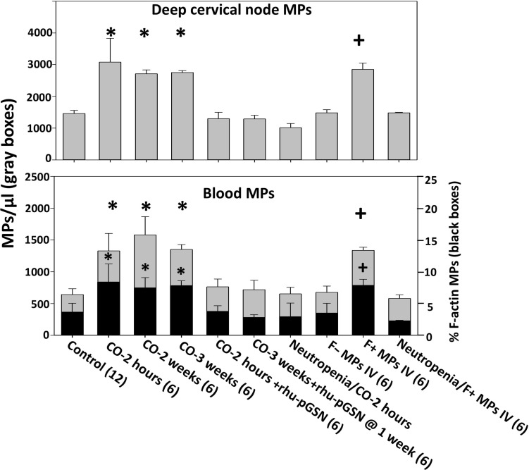
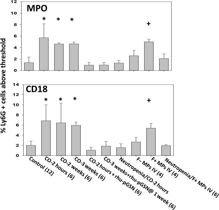
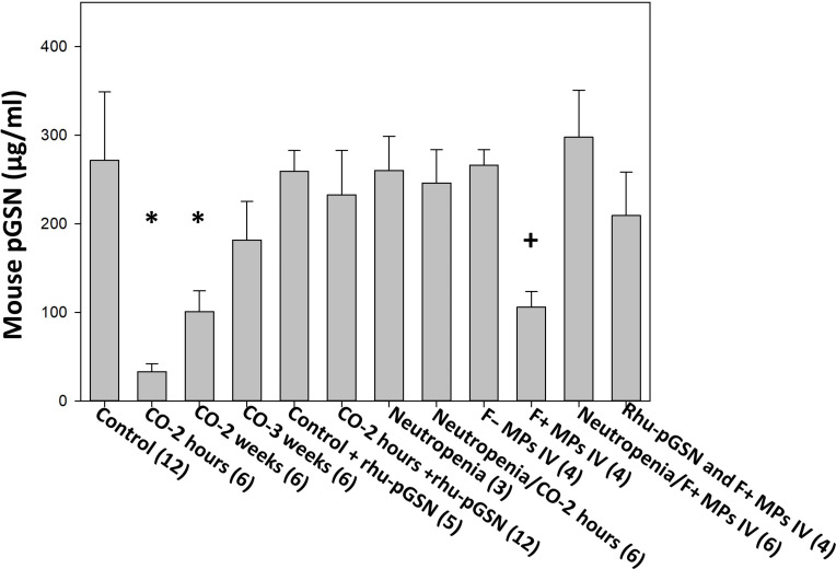
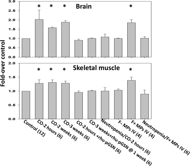
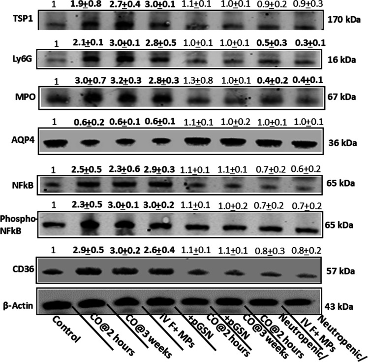
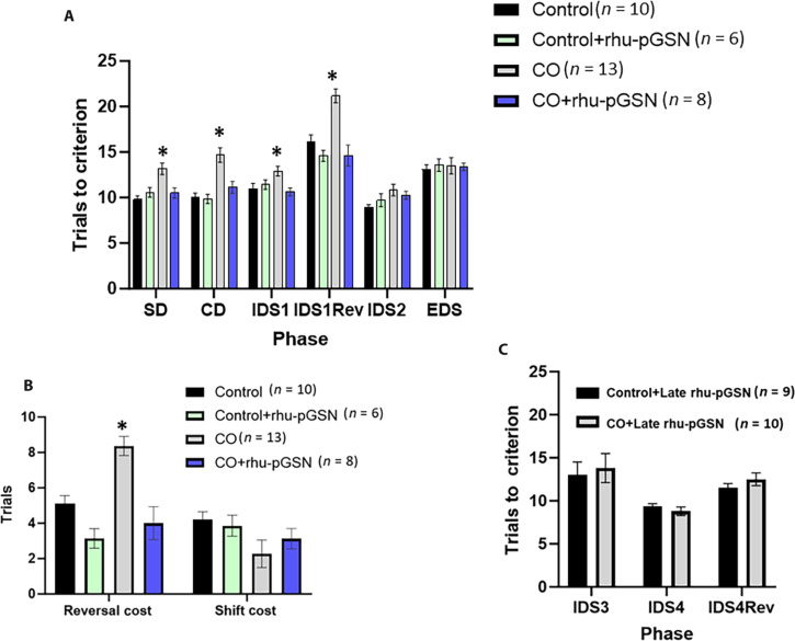
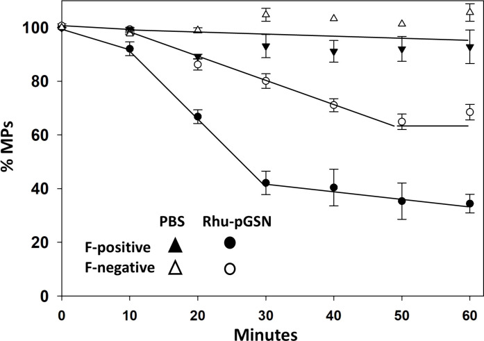
The mechanism for neurological deficits from carbon monoxide (CO) poisoning is unclear. In a series of 150 patients with CO poisoning, we found marked elevations of blood-borne inflammatory filamentous (F-) actin-coated microparticles (MPs), neutrophil activation, and a 90% reduction in the normal level of plasma gelsolin (pGSN), a protein capable of lysing F-actin-coated MPs. This led to studies in a murine model where the same events occur and cause neuroinflammation with cognitive dysfunction. All events are recapitulated when F-actin MPs are injected intravenously, which establishes a blood-to-brain-to-blood inflammatory cycle that persists for weeks. All changes, including cognitive dysfunction, can be abrogated by an injection of human recombinant pGSN within 2 weeks after CO poisoning. These findings demonstrate that CO-induced neurological injury has an inflammatory etiology. Because of MP-mediated communications between the brain and systemic circulation, CO-induced cognitive deficits may be reversible with a pharmaceutical intervention.


Figures

Fig. 1.
Fig. 1.. Deep cervical lymph node and blood microparticles (MPs).
The total number of MPs in deep cervical nodes and blood (gray boxes) and % of blood MPs expressing F-actin (black boxes) are shown. The data represent mean ± SD (n for each group shown along the abscissa), where *P < 0.001 vs. control (ANOVA) and +P < 0.001 for those injected with F-positive MPs versus F-negative MPs (t test). Subgroups of MPs expressing protein markers are shown in table S1 (A and B). Groups include control; mice exposed to CO and euthanized 2 hours later, 2 weeks later, and 3 weeks later; mice exposed to CO and immediately injected with a sterile solution of rhu-pGSN (38.4 mg/ml) at a dose of 27 mg/kg intravenously and euthanized 2 hours later; mice exposed to CO, injected with rhu-pGSN immediately after the CO exposure, and euthanized 3 weeks later; mice rendered neutropenic, exposed to CO, and euthanized 2 hours later; naïve mice injected with 60,000 MPs in 200 μl of sterile PBS that were F-actin negative (F− MPs) or F-actin positive (F+ MPs); and neutropenic mice injected with F-actin–positive MPs.
Fig. 2.
Fig. 2.. Neutrophil activation.
Neutrophil activation was assessed by flow cytometry as the % of Ly6G positive cells expressing myeloperoxidase (MPO) and CD18 on the membrane surface above a threshold concentration present on control cells. Mouse groups and data are as described in Fig. 1. *P < 0.001 vs. control (ANOVA) and +P < 0.001 for those injected with F-positive MPs versus F-negative MPs (t test).
Fig. 3.
Fig. 3.. Plasma gelsolin values.
The data indicators and mouse groups are as described in Fig. 1. Additional groups in the figure include control mice injected with rhu-pGSN at 27 mg/kg intravenously and euthanized 2 hours later, neutropenic mice not exposed to CO, and mice injected with 60,000 F-actin MPs in 200 μl of sterile PBS where the MPs were first incubated for 15 min with rhu-pGSN to cause particle lysis (20). The rhu-pGSN dose was the same as in other groups (27 mg/kg mouse) so that for an average 20-g mouse, the F-actin MPs had been incubated with rhu-pGSN at a concentration of ~2.7 μg/μl. *P < 0.001 vs. control (ANOVA) and +P < 0.001 for those injected with F-positive MPs versus F-negative MPs (t test).
Fig. 4.
Fig. 4.. Murine vascular leakage of 2 × 10⁶ Da rhodamine-labeled dextran.
Extravasation of dextran in brain and leg skeletal muscle was evaluated as described in Materials and Methods. Mouse groups are as described in Fig. 1. Data are fold difference in rhodamine-dextran/mg tissue protein (mean ± SD) versus the values in control mice processed concurrently with each experimental group. *P < 0.001 vs. control (ANOVA) and +P < 0.001 for those injected with F-positive MPs versus F-negative MPs (t test).
Fig. 5.
Fig. 5.. Neuroinflammation assessed by Western blot.
A representative Western blot of brain homogenates is shown for mouse groups as described in Fig. 1 (not shown are data for 2 weeks post-CO and mice injected with F-actin–negative MPs as values for these mice were not significantly different from control) where numbers above each band indicate mean ± SD (n = 6 mouse brains per lane) band densities relative to β-actin of control samples from replicate studies. Bold numbers indicate values statistically significant from control (P ≤ 0.01 ANOVA). Blots were probed for NF-κB, p65-phosphorylated NF-κB (pNF-κB), myeloperoxidase (MPO), thrombospondin-1 (TSP), Ly6G, and CD36 proteins.
Fig. 6.
Fig. 6.. Attentional set shift scores for control and CO-exposed mice.
(A) Trials to criterion for progressive tasks. Where indicated, rhu-pGSN injection occurred immediately after CO exposure and control mice injected at the same time. The CO group demonstrated differences on each of the first four phases, the smallest value F(3,33) = 4.09, P < 0.01 (ANOVA). The CO mice significantly differed from control mice on each of these phases, P < 0.05 (*). The trials to criterion to complete each phase was significant, F(5,165) = 53.55, P < 0.001 (RM-ANOVA) as a phase × exposure interaction, F(5,165) = 2.91, P < 0.015, and phase × treatment interaction, F(5,165) = 5.71, P < 0.001. The main effects of CO, F(1,33) = 22.89, P < 0.001, rhu-pGSN treatment, F(1,33) = 15.53, P < 0.001, and the interaction, F(1,33) = 19.10, P < 0.001, were significant. (B) Reversal and extradimensional shift cost scores. No significant differences were detected on the extradimensional shift cost, but CO-exposed mice were significantly less likely to demonstrate an attentional set formation as confirmed by the χ2 statistic. Values are mean ± SD, * indicates P < 0.05 versus control. (C) Effect of late rhu-pGSN administration. After tasks were performed as in (B), mice were injected with rhu-pGSN so that during the third week, repetitive IDS testing (IDS 3 and 4) and reversal tasks were assessed. No significant differences between CO-exposed and control mice were found.
Fig. 7.
Fig. 7.. Lysis of human MPs by rhu-pGSN.
Suspensions of MPs from four patients with CO poisoning were first separated into those expressing F-actin (F-positive) and those not expressing F-actin (F-negative). Suspension of 1200 MPs in 250 μl of PBS was then incubated with 19 μg of rhu-pGSN (76 μg/ml) or PBS and samples were taken at intervals for particle counting. Data show mean ± SD MPs/μl at up to 1 hour of incubation.

Similar articles

References

    1. Raub J. A., Mathieu-Nolf M., Hampson N. B., Thom S. R., Carbon monoxide poisoning—A public health perspective. Toxicology 145, 1–14 (2000). - PubMed
    1. Mattiuzzi C., Lippi G., Worldwide epidemiology of carbon monoxide poisoning. Hum. Exp. Toxicol. 39, 387–392 (2020). - PubMed
    1. Centers for Disease Control and Prevention (CDC) , Carbon monoxide-related deaths—United States, 1999-2004. MMWR Morb. Mortal. Wkly Rep. 56, 1309–1312 (2007). - PubMed
    1. Shin M., Bronstein A. C., Glidden E., Malone M., Chang A., Law R., Boehmer T. K., Strosnider H., Yip F., Morbidity and mortality of unintentional carbon monoxide poisoning: United States 2005 to 2018. Ann. Emerg. Med. 81, 309–317 (2023). - PMC - PubMed
    1. Hampson N. B., Weaver L. K., Carbon monoxide poisoning: A new incidence for an old disease. Undersea Hyperb. Med. 34, 163–168 (2007). - PubMed