. 2025 Feb 27;68(4):4217-4236.
doi: 10.1021/acs.jmedchem.4c01998. Epub 2025 Feb 7.

MTA-Cooperative PRMT5 Inhibitors: Mechanism Switching Through Structure-Based Design

Affiliations

MTA-Cooperative PRMT5 Inhibitors: Mechanism Switching Through Structure-Based Design

Kevin M Cottrell et al. J Med Chem. .

Abstract

Deletion of the MTAP gene leads to accumulation of the substrate of the MTAP protein, methylthioadenosine (MTA). MTA binds PRMT5 competitively with S-adenosyl-l-methionine (SAM), and selective inhibition of the PRMT5•MTA complex relative to the PRMT5•SAM complex can lead to selective killing of cancer cells with MTAP deletion. Herein, we describe the discovery of novel compounds using structure-based drug design to switch the mechanism of binding of known, SAM-cooperative PRMT5 inhibitors to an MTA-cooperative binding mechanism by occupying the portion of the SAM binding pocket in PRMT5 that is unoccupied when MTA is bound and hydrogen bonding to Arg368, thereby allowing them to selectively target MTAP-deleted cancer cells.


Conflict of interest statement

The authors declare no competing financial interest.

Figures

Figure 1
Figure 1
Chemical structures of the clinical MTA-cooperative PRMT5 inhibitors TNG908, TNG462, MRTX1719 (BMS-986504), and AMG 193.
Figure 2
Figure 2
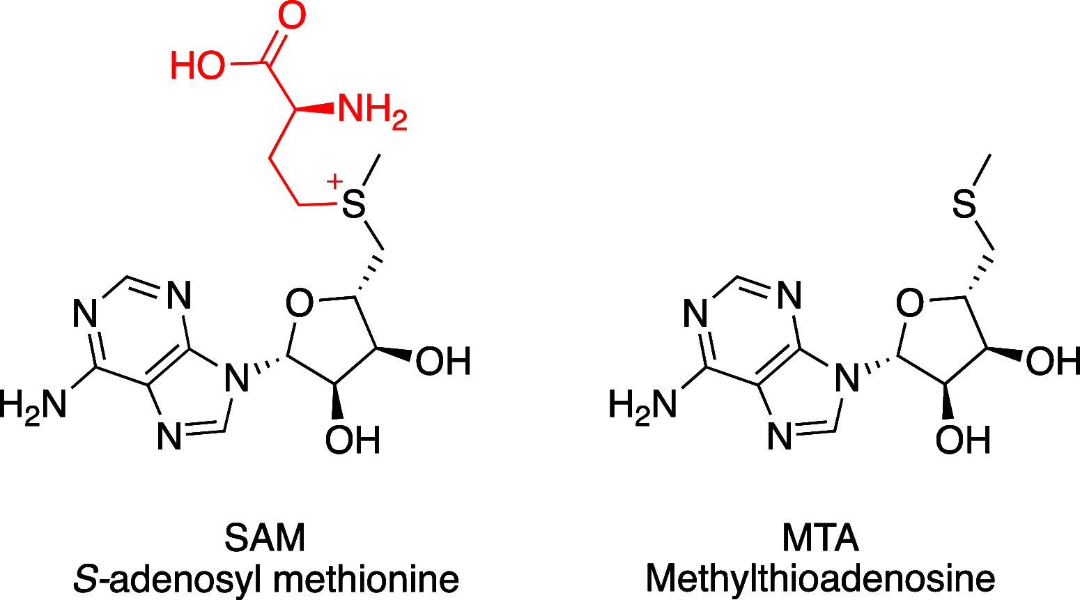
Chemical structures of S-adenosyl methionine (SAM) and methylthioadenosine (MTA).
Figure 3
Figure 3
X-ray crystal structure of GSK3326595 (pemrametostat; blue, PDB9MGL) with PRMT5•MTA reveals a small pocket adjacent to the 6-position of the tetrahydroisoquinoline relative to the structure with SAM.
Figure 4
Figure 4
X-ray crystal structure of 24 (green, PDB entry 9MGM) with PRMT5•MTA. The crystal structure shows the ether extension reaching into the SAM binding pocket and making a hydrogen bond from the pyrazole N to the backbone NH of Arg368.
Figure 5
Figure 5
X-ray crystal structure of 41 (purple, PDB entry 9MGN) bound to PRMT5•MTA with pyrazole forming two hydrogen bonds to Arg368 and Glu328.
Figure 6
Figure 6
Cyclization of the central chain of A leads to B as a hypothesis for design that led to 46a.
Figure 7
Figure 7
X-ray crystal structures of 46a (magenta, PDB 9MGP) overlaid on 24 (green) showing the methylpyrazole subgroup flipped in the binding pocket but each still engaging Arg368 in a hydrogen bond.
Figure 8
Figure 8
X-ray crystal structure overlays of 47 (yellow, PDB entry 9MGQ) in PRMT5•sinefungin and 24 (green) in PRMT5•MTA suggest that 7-substitution on 47 provides the best opportunity to access the SAM pocket to achieve MTA cooperativity. Only the protein and MTA when bound with 24 is depicted.
Figure 9
Figure 9
In cell western SDMA assay data for 54 are compared to GSK3226595 in MTAP-del and MTAP WT cells. 54 is equipotent to GSK3226595 in MTAP-del cells but is selective relative to MTAP WT cells.
Figure 10
Figure 10
X-ray crystal structure of 51 (blue, PDB entry 9MGR) in PRMT5•MTA.
Figure 11
Figure 11
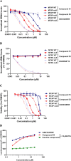
In vitro data: (A) SDMA in cell western (ICW) PD data for HAP1MTAP-null vs MTAP WT cells for 51 and 53. and (B) ADMA ICW PD data for HAP1MTAP-null vs MTAP WT cells for 51 and 53. GSK3368712 is a type I PRMT inhibitor and included as an assay control. (C) Cellular viability data in HAP1 MTAP-null vs MTAP WT cells for 51 and 53. (D) Thermostability target engagement in HAP1 MTAP-null cells engineered to stably express an exogenous NanoLuc-PRMT5 protein for 53.
Scheme 1
Scheme 1. Synthesis of Compound 24
Reagents and conditions: (a) Boc2O, DMAP, MTBE. (b) Cyclobutylamine, K2CO3, DMF, 100 °C. (c) HCl, EtOAc, 50 °C. (d) (S)-1-Amino-3-chloropropan-2-ol hydrochloride, HATU, DIPEA, DCM, DMF. (e) PPh3, DIAD, toluene, 20–90 °C. (f) 4 M HCl, dioxane. (g) DIPEA, NaI, DBU, ACN.
Scheme 2
Scheme 2. Synthesis of Compound 53
Reagents and conditions: (a) methoxymethyl chloride, cesium carbonate, DMF. (b) DIBAL-H, MeOH, DCM. (c) MeNO2, (1R, 2R)-N1,N2-bis[(4-chlorophenyl)methyl]cyclohexane-1,2-diamine copper(II) acetate hydrate, EtOH, MTBE. (d) 10% Pd/C, MeOH. (e) TEA, DCM. (f) piperidine, TEA, ACN. (g) LiOH•H2O, MeOH, THF, H2O. (h) AcCl, TEA, ACN. (i) HATU, TEA, DMF. (j) 4 M HCl, dioxane. (k) NaHCO3, Boc2O, THF, H2O. (l) 5-(chloromethyl)-4-methyloxazole, Cs2CO3, DMF. (m) 4 M HCl, dioxane.
Scheme 3
Scheme 3. Synthesis of Compounds 45a and 45b
Reagents and conditions: (a) HCl, EtOAc, 50 °C. (b) KOH, MeOH, 0 °C. (c) I2, MeOH, 0–20 °C. (d) p-TsOH•H2O, toluene. (e) NaBH(OAc)3, THF. (f) 4 N HCl, EtOAc, DCM. (g) HATU, DIPEA, DCM, DMF. (h) Chiral SFC separation.
Scheme 4
Scheme 4. Synthesis of Compounds 46a and 46b
Reagents and conditions: (a) mCPBA, DCM. (b) CO (50 psi), Pd(dppf)Cl2, TEA, TES, DMF, 80 °C. (c) nBuLi, hexanes, THF, −78 °C. (d) THF, −78 °C–20 °C. (e) Pd/C, H2 (15 psi), HCl/H2O, tBuOH, 70 °C. (f) 75, TEA, EtOH, 100 °C. (g) TFA, DCM. (h) 70, HATU, DIPEA, DCM, DMF. (i) Chiral SFC separation.

References

    1. Stopa N.; Krebs J. E.; Shechter D. The PRMT5 Arginine Methyltransferase: Many Roles in Development, Cancer and Beyond. Cell. Mol. Life Sci. 2015, 72 (11), 2041–2059. 10.1007/s00018-015-1847-9. - DOI - PMC - PubMed
    1. Blanc R. S.; Richard S. Arginine Methylation: The Coming of Age. Mol. Cell 2017, 65 (1), 8–24. 10.1016/j.molcel.2016.11.003. - DOI - PubMed
    1. Musiani D.; Bok J.; Massignani E.; Wu L.; Tabaglio T.; Ippolito M. R.; Cuomo A.; Ozbek U.; Zorgati H.; Ghoshdastider U.; Robinson R. C.; Guccione E.; Bonaldi T. Proteomics Profiling of Arginine Methylation Defines PRMT5 Substrate Specificity. Sci. Signaling 2019, 12 (575), eaat838810.1126/scisignal.aat8388. - DOI - PubMed
    1. Chan-Penebre E.; Kuplast K. G.; Majer C. R.; Boriack-Sjodin P. A.; Wigle T. J.; Johnston L. D.; Rioux N.; Munchhof M. J.; Jin L.; Jacques S. L.; West K. A.; Lingaraj T.; Stickland K.; Ribich S. A.; Raimondi A.; Scott M. P.; Waters N. J.; Pollock R. M.; Smith J. J.; Barbash O.; Pappalardi M.; Ho T. F.; Nurse K.; Oza K. P.; Gallagher K. T.; Kruger R.; Moyer M. P.; Copeland R. A.; Chesworth R.; Duncan K. W. A Selective Inhibitor of PRMT5 with in Vivo and in Vitro Potency in MCL Models. Nat. Chem. Biol. 2015, 11 (6), 432–437. 10.1038/nchembio.1810. - DOI - PubMed
    1. Brehmer D.; Beke L.; Wu T.; Millar H. J.; Moy C.; Sun W.; Mannens G.; Pande V.; Boeckx A.; van Heerde E.; et al. Discovery and Pharmacological Characterization of JNJ-64619178, a Novel Small Molecule Inhibitor of PRMT5 with Potent Anti-Tumor Activity. Mol. Cancer Ther. 2021, 20 (12), 2317–2328. 10.1158/1535-7163.mct-21-0367. - DOI - PMC - PubMed

MeSH terms

LinkOut - more resources