. 2025 Feb 7;16(1):1438.
doi: 10.1038/s41467-025-56737-6.

Aspirin inhibits proteasomal degradation and promotes α-synuclein aggregate clearance through K63 ubiquitination

Affiliations

Aspirin inhibits proteasomal degradation and promotes α-synuclein aggregate clearance through K63 ubiquitination

Jing Gao et al. Nat Commun. .

Abstract

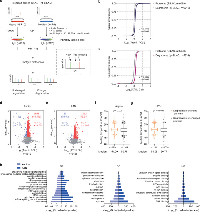
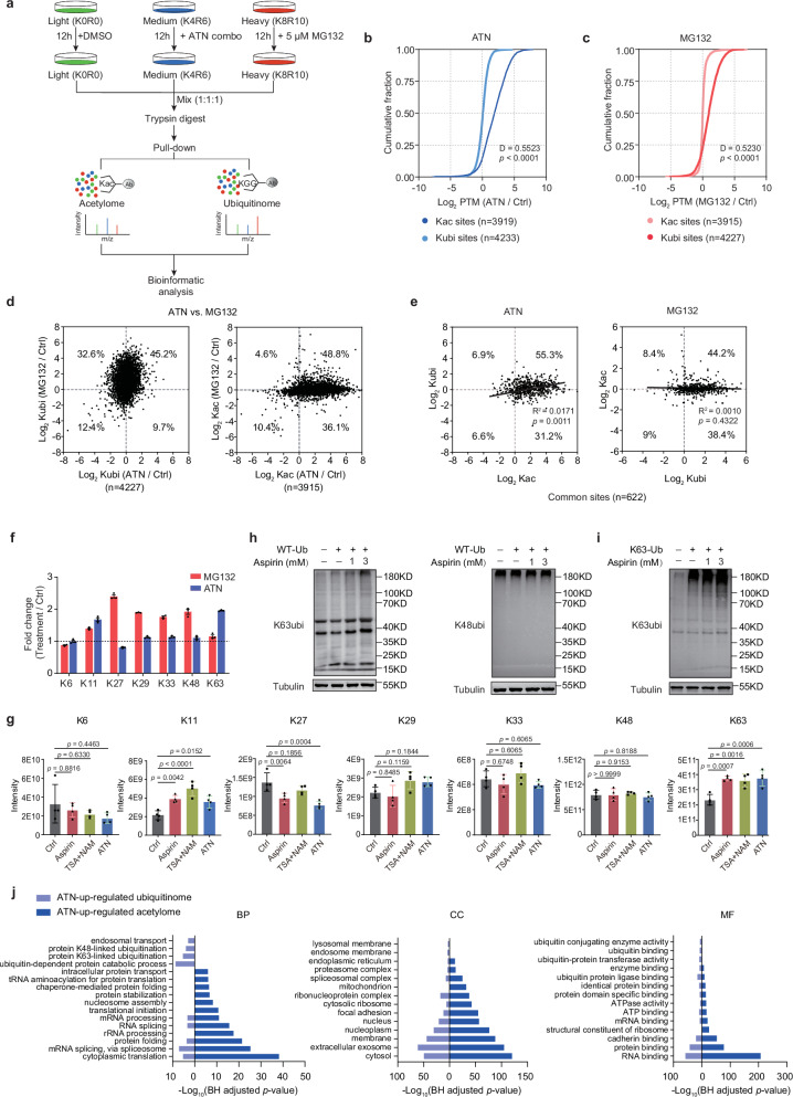
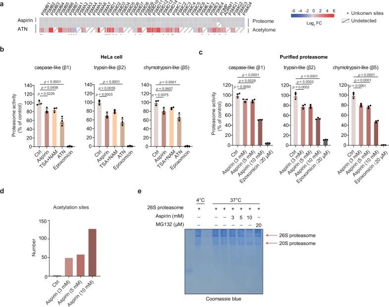
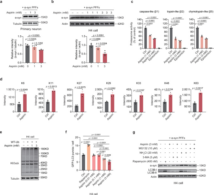
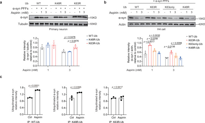
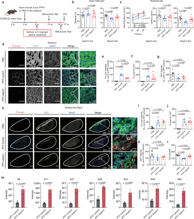
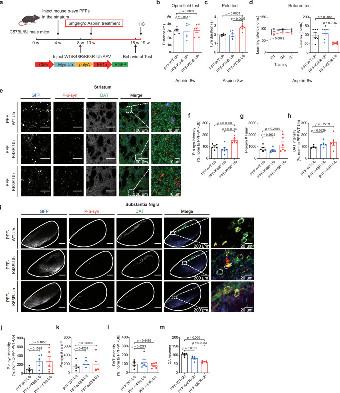
Aspirin is a potent lysine acetylation inducer, but its impact on lysine ubiquitination and ubiquitination-directed protein degradation is unclear. Herein, we develop the reversed-pulsed-SILAC strategy to systematically profile protein degradome in response to aspirin. By integrating degradome, acetylome, and ubiquitinome analyses, we show that aspirin impairs proteasome activity to inhibit proteasomal degradation, rather than directly suppressing lysine ubiquitination. Interestingly, aspirin increases lysosomal degradation-implicated K63-linked ubiquitination. Accordingly, using the major pathological protein of Parkinson's disease (PD), α-synuclein (α-syn), as an example of protein aggregates, we find that aspirin is able to reduce α-syn in cultured cells, neurons, and PD model mice with rescued locomotor ability. We further reveal that the α-syn aggregate clearance induced by aspirin is K63-ubiquitination dependent in both cells and PD mice. These findings suggest two complementary mechanisms by which aspirin regulates the degradation of soluble and insoluble proteins, providing insights into its diverse pharmacological effects that can aid in future drug development efforts.


Conflict of interest statement

Competing interests: Y.Z., J.G. and K.H. have filed a patent application related to the use of aspirin or its derivatives for treating diseases associated with abnormal aggregation of α-syn. All other authors declare no competing interests. Ethics: All animal experimental procedures were reviewed and approved by the Institutional Animal Care and Use Committee at the Interdisciplinary Research Center on Biology and Chemistry, Chinese Academy of Sciences, and are in accordance with the Guide for the Care and Use of Laboratory Animals of the Chinese Academy of Sciences.

Figures

Fig. 1
Fig. 1. rp-SILAC strategy reveals aspirin-mediated inhibition of protein degradation in HeLa cells.
a Experimental scheme of the rp-SILAC strategy. This strategy utilizes quantification between the MS signals representing the pre-existing proteins to analyze degradation. The treatment with aspirin (b) or ATN combo (c) leads to the suppression of protein degradation. Aspirin or ATN combo treatment decreases global degradome slightly, but does not affect proteins’ abundance significantly. The distance (“D”) and p-value were calculated using the two-tailed Kolmogorov-Smirnov test (Proteome: n = 3 independent experiments; Degradome: n = 4 independent experiments). Volcano plots display the change in degradome caused by aspirin (d) or ATN combo (e) treatment. Proteins regulated by degradation (p < 0.05) are highlighted in red (FC > 1) or blue (FC < 1) dots, and the number of these proteins is displayed on the plots. Statistical tests were performed using one-sample two-tailed t-test with Benjamini-Hochberg multiple test correction. Melting temperature of degradation-changed proteins (p < 0.05) and those unchanged (p ≥ 0.05) with aspirin (f) or ATN combo (g) treatment. Boxes represent the interquartile range (25th to 75th percentile) for melting temperature, and whiskers extend from the 2.5th to the 97.5th percentile of the data. Two-tailed Mann-Whitney U test was used to analyze the significance of differences between groups (Aspirin: n = 2214 and 1364; ATN: n = 2281 and 1059). h Gene Ontology analysis of degradation-changed proteins. Fisher’s Exact test, with Benjamini-Hochberg adjusted p-values, is used to measure gene enrichment in annotation terms. Source data are provided as a Source Data file.
Fig. 2
Fig. 2. Aspirin-induced lysine acetylation does not directly interfere with lysine ubiquitination in HeLa cells.
a Quantitative experimental scheme for elucidating crosstalk between lysine acetylome and ubiquitinome. Triple SILAC-based quantitative proteomics was integrated with antibody-based acetylation and ubiquitination enrichment. Lysine acetylation and ubiquitination do not have a significant effect on each other. Cumulative distributions show that ATN combo treatment significantly increases acetylation level but does not affect ubiquitination (b). Similarly, MG132 treatment only changes ubiquitination and not acetylation (c). The distance (“D”) and p-value were calculated using the two-tailed Kolmogorov-Smirnov test (n = 3 independent experiments). d Comparisons of individual ubiquitination (left) and acetylation (right) sites between ATN and MG132 treatments. e Relationship between co-occurring lysine acetylation and ubiquitination. The Pearson correlation coefficients show that no clear positive or negative correlations were observed between these two modifications. f ATN combo or MG132 treatment regulates ubiquitin linkages. Peptides MQIFVK(GG)TLTGK, TLTGK(GG)TITLEVEPSDTIENVK, TITLEVEPSDTIENVK(GG)AK, AK(GG)IQDK, IQDK(GG)EGIPPDQQR, LIFAGK(GG)QLEDGR, and TLSDYNIQK(GG)ESTLHLVLR are used to represent K6, K11, K27, K29, K33, K48, and K63 ubiquitination linkages, respectively. The quantification MS data retrieved from the triple-SILAC ubiquitinome experiment are shown (n = 3 independent experiments. data presented as mean ± SEM). g The effect of aspirin, TSA, and NAM, and their combination, on ubiquitin linkages. The quantification MS data were obtained from a separate label-free experiment using HeLa cells. The signature peptides representing seven types of ubiquitin linkages were used for quantification (n = 4 independent experiments, One-way ANOVA with Dunnett’s multiple comparisons test, data presented as mean ± SEM). h The impact of aspirin on K63 ubiquitination (left) and K48 ubiquitination (right) in HeLa cells. Recombinant wild-type ubiquitin was introduced to enhance quantification performance. Three independent experiments were conducted. i Aspirin induces K63 ubiquitination in HeLa cells. Recombinant K63-only ubiquitin was introduced to improve quantification performance on K63 specific ubiquitination linkage. Three independent experiments were conducted. j Gene Ontology analysis of proteins containing up-regulated (FC > 1.5) acetylated and ubiquitinated sites with ATN combo treatment. Fisher’s Exact test, with Benjamini-Hochberg adjusted p-values, is used to measure gene enrichment in annotation terms. Source data are provided as a Source Data file.
Fig. 3
Fig. 3. Aspirin impairs proteolytic activity of proteasome.
a Aspirin or ATN combo treatment induces acetylation on many proteasome subunits. The protein abundances of these proteasome components remain unchanged upon aspirin or ATN combo treatment. 25 acetylation sites were previously unknown and are labeled with asterisks. Aspirin decreases proteolytic activities of three major proteasomal hydrolases from HeLa cell extracts (b) or purified proteasome (c). Epoxomicin is a known potent and selective inhibitor of the proteasome. HeLa cells were treated with 3 mM aspirin, 10 μM TSA, and 10 mM NAM. After incubating proteasome with fluorogenic peptide substrates at 37 °C for 60 min, the fluorescence intensity was recorded (n = 3 independent experiments, One-way ANOVA with Dunnett’s multiple comparisons test, data presented as mean ± SEM). d The number of acetylation sites on the proteasome induced by aspirin in vitro. e The effect of aspirin on the integrity of 26S proteasome. 26S proteasome remains intact under low concentration (3 or 5 mM) of aspirin, but undergoes partial disassembly under high concentration (10 mM) in vitro. 6% native page gels were used to separate the capped proteasome (26S) from 20S proteasome. Source data are provided as a Source Data file.
Fig. 4
Fig. 4. Aspirin promotes clearance of α-syn aggregates in neurons and neuroglioma H4 cells.
Aspirin treatment decreases α-syn aggregates in mouse primary neurons (a) and neuroglioma H4 cells (b). Shown are representative western blot (top) and quantitative results (bottom) (n = 3 independent experiments, One way ANOVA with Tukey’s multiple comparisons test, data presented as mean ± SEM). c Proteolytic activities of three major proteasomal hydrolases from H4 cell extracts decrease upon aspirin treatment (n = 3 independent experiments, One way ANOVA with Dunnet’s multiple comparisons test, data presented as mean ± SEM). d Aspirin treatment alters ubiquitin linkages in H4 cells. Shown are the quantification MS data retrieved from a label-free experiment using H4 cells treated with 3 mM aspirin for 18 h. The quantification was based on the signature peptides representing seven types of ubiquitin linkages. (n = 3 independent experiments, unpaired two-tailed t-test, data presented as mean ± SEM). e Aspirin induces K63 ubiquitination in H4 cells. Recombinant wild-type ubiquitin was introduced to enhance quantification performance. Three independent experiments were conducted. f Quantification of GFP-LC3 puncta number per cell after treatment with varied concentrations of aspirin in H4-GFP-LC3 cells. At least 1000 cells per condition in an independent experiment were quantified (n = 5 independent experiments, One way ANOVA with Tukey’s multiple comparisons test, data presented as mean ± SEM). g Aspirin’s reduction of α-syn is lysosome dependent. Inhibiting lysosome activity with NH4Cl significantly diminishes aspirin’s effectiveness in α-syn clearance. Three independent experiments were conducted. Source data are provided as a Source Data file.
Fig. 5
Fig. 5. Aspirin mediates the degradation of α-syn aggregates through a K63 ubiquitination-dependent endosomal pathway.
K63 ubiquitination is the key signaling mediator for the degradation of α-syn aggregates in primary neurons (a) and H4 cells (b). Shown are representative western blot (top) and corresponding quantitative results (bottom). The overexpression of K63R ubiquitin variant in both primary neurons and H4 cells diminishes the aspirin-induced clearance of α-syn aggregates (n = 3 independent experiments, One-way ANOVA with Tukey’s multiple comparisons test, data presented as mean ± SEM). c Aspirin induces K63-linked ubiquitination on α-syn. Ubiquitinated proteins were immunoprecipitated using an anti-myc pull-down, and α-syn levels were measured by mass spectrometry (n = 3 independent experiments, unpaired two-tailed t-test, data presented as mean ± SEM). Source data are provided as a Source Data file.
Fig. 6
Fig. 6. Aspirin promotes clearance of α-syn aggregates in a PD mouse model.
a Experimental scheme for establishing a PD mouse model and aspirin treatment. 10, 11, and 12 mice were used for the PBS, the PFF-control, and the PFF-aspirin treatments, respectively. The figure was created using BioRender.com. Open field test (b) and rotarod test (c) for PD mice. These behavioral tests were repeated twice during the aspirin administration. The results of the rotarod test were normalized to the batch-specific PBS mice in each independent experimental batch. Each dot represents a mouse (One way ANOVA with FDR correction for multiple comparisons, data presented as mean ± SEM). d Aspirin treatment decreases α-syn aggregates in the striatum. Representative immunofluorescence images (scale bar, 100 μm) and zoom-in merged images (scale bar, 10 μm) are shown. Quantitative results are shown in eg, and the dot represents the mouse brain. Aspirin treatment reduces the intensity (e) and density (f) of α-syn aggregates in the striatum, which is represented by p-α-syn (n = 5 or 6, One way ANOVA with FDR correction for multiple comparisons, data presented as mean ± SEM). g DA neurons in the striatum with aspirin treatment (n = 5 or 6, One way ANOVA with Tukey’s multiple comparisons test). h Aspirin treatment increases the number of DA neurons in the substantia nigra. Representative immunofluorescence images (scale bar, 200 μm) and zoom-in merged images (scale bar, 20 μm) are shown. Quantitative results are shown in il, and the dot represents the mouse brain. Intensity (i) and density (j) of α-syn aggregates in the substantia nigra with aspirin treatment (n = 6, One way ANOVA with FDR correction for multiple comparisons, data presented as mean ± SEM). DA neurons in the substantia nigra with aspirin treatment (n = 6, One way ANOVA with FDR correction for multiple comparisons, data presented as mean ± SEM). m Aspirin treatment regulates ubiquitin linkages in mouse brains. The signature peptides representing seven types of ubiquitin linkages were used for quantification (n = 3, unpaired two-tailed t-test, data presented as mean ± SEM). Source data are provided as a Source Data file.
Fig. 7
Fig. 7. Aspirin-induced α-syn clearance is dependent on K63 ubiquitination in PD mice.
a Experimental scheme for aspirin treatment in a PD mouse model overexpressing different ubiquitin variants. AAV vectors expressing three ubiquitin variants (WT, K48R, K63R) were introduced to PD mice. 6 mice were used for each of the PFF-WT-Ub, the PFF-K48R-Ub, and the PFF-K63R-Ub treatments, respectively. The figure was created using BioRender.com. Open field test (b), pole test (c) and rotarod test (d) for PD mice with different ubiquitin variants. Each dot represents a mouse (n = 6, One-way ANOVA with FDR correction for multiple comparisons, data presented as mean ± SEM). e The K63R ubiquitin significantly weakens the clearance effect of p-α-syn caused by aspirin in the striatum. Representative immunofluorescence images of the striatum (scale bar, 100 μm) are shown. Zoom-in merged images are shown on the right (scale bar, 10 μm). Quantitative results are shown in fh, and the dot represents the mouse brain. Impact of different ubiquitin variants, when treated with aspirin, on p-α-syn intensity (f) and density (g), as well as DAT intensity (h) in the striatum (n = 5 or 6, One way ANOVA with FDR correction for multiple comparisons, data expressed as mean ± SEM). i The K63R ubiquitin significantly decreases the number of DA neurons in the substantia nigra. Shown are representative immunofluorescence images of the substantia nigra (scale bar, 200 μm). Zoom-in of the merged images are shown on the right (scale bar, 20 μm). Quantitative results are shown in jm, and the dot represents the mouse brain. Impact of different ubiquitin variants, when treated with aspirin, on p-α-syn intensity (j) and density (k), as well as DAT intensity (l) and DA neuron number (m) in the substantia nigra. The number of DA neurons was determined by counting DAT-positive cells in the substantia nigra (n = 4, 5 or 6, One way ANOVA with FDR correction for multiple comparisons, data expressed as mean ± SEM). Source data are provided as a Source Data file.

Similar articles

Cited by

References

    1. Prasad, K. N., Wu, M. & Bondy, S. C. Telomere shortening during aging: Attenuation by antioxidants and anti-inflammatory agents. Mech. Ageing Dev.164, 61–66 (2017). - PubMed
    1. Flossmann, E. & Rothwell, P. M. Effect of aspirin on long-term risk of colorectal cancer: consistent evidence from randomised and observational studies. Lancet369, 1603–1613 (2007). - PubMed
    1. Ayyadevara, S. et al. Aspirin inhibits oxidant stress, reduces age-associated functional declines, and extends lifespan of Caenorhabditis elegans. Antioxid. Redox Signal18, 481–490 (2013). - PubMed
    1. Green, G. A. Understanding NSAIDs: from aspirin to COX-2. Clin. Cornerstone3, 50–60 (2001). - PubMed
    1. Guo, L., Gao, J., Gao, Y., Zhu, Z. & Zhang, Y. Aspirin reshapes acetylomes in inflammatory and cancer cells via CoA-dependent and CoA-independent pathways. J. Proteome19, 962–972 (2020). - PubMed

MeSH terms

LinkOut - more resources