. 2025 Feb 7;22(1):33.
doi: 10.1186/s12974-025-03356-z.

Inhibition of tumour necrosis factor alpha by Etanercept attenuates Shiga toxin-induced brain pathology

Affiliations

Inhibition of tumour necrosis factor alpha by Etanercept attenuates Shiga toxin-induced brain pathology

Robin Christ et al. J Neuroinflammation. .

Abstract

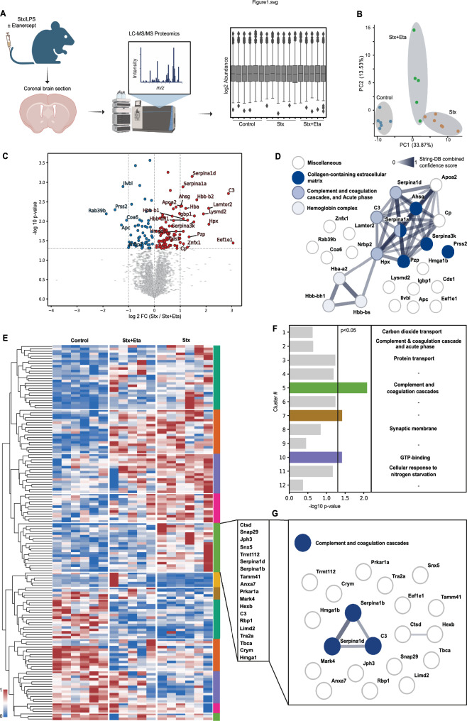
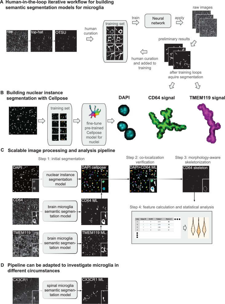
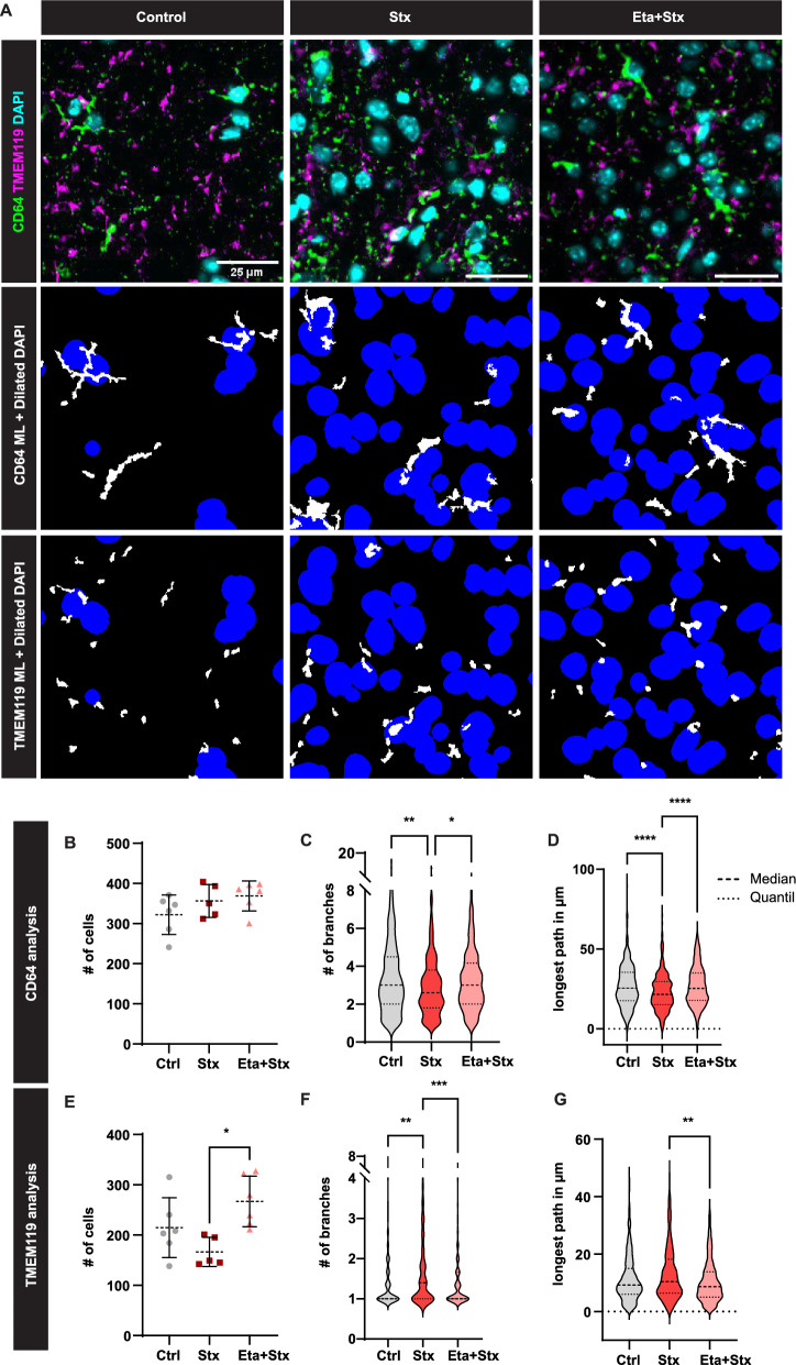
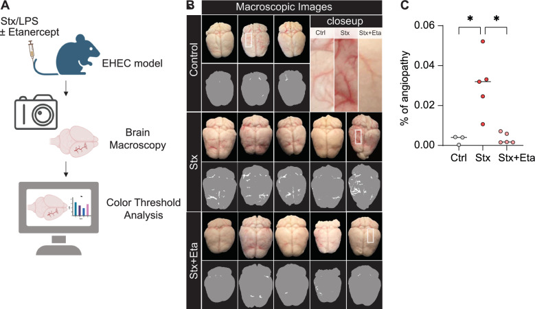
Infection with enterohemorrhagic E. coli (EHEC) causes severe changes in the brain leading to angiopathy, encephalopathy and microglial activation. In this study, we investigated the role of tumour necrosis factor alpha (TNF-α) for microglial activation and brain pathology using a preclinical mouse model of EHEC infection. LC-MS/MS proteomics of mice injected with a combination of Shiga toxin (Stx) and lipopolysaccharide (LPS) revealed extensive alterations of the brain proteome, in particular enrichment of pathways involved in complement activation and coagulation cascades. Inhibition of TNF-α by the drug Etanercept strongly mitigated these changes, particularly within the complement pathway, suggesting TNF-α-dependent vasodilation and endothelial injury. Analysis of microglial populations using a novel human-in-the-loop deep learning algorithm for the segmentation of microscopic imaging data indicated specific morphological changes, which were reduced to healthy condition after inhibition of TNF-α. Moreover, the Stx/LPS-mediated angiopathy was significantly attenuated by inhibition of TNF-α. Overall, our findings elucidate the critical role of TNF-α in EHEC-induced brain pathology and highlight a potential therapeutic target for mitigating neuroinflammation, microglial activation and injury associated with EHEC infection.


Conflict of interest statement

Declarations. Ethics approval and consent to participate: The animal experiments were approved by the local animal review board of the government (Bezirksregierung Köln, Landesamt für Natur, Umwelt und Verbraucherschutz NRW in Recklinghausen, Germany). The Ethical Subcommittee in Life and Health Science (SECVS; 018/2019, University of Minho) and the ORBEA (EM/ICVS-I3Bs_017-2021) approved all experiments. Competing interests: The authors declare no competing interests.

Figures

Fig. 1
Fig. 1
Inhibition of TNF-α reduces the activation of the complement and coagulation cascade. A Schematic illustration of the measurement of coronal brain section of mice by liquid chromatography mass spectrometry (LC–MS/MS). Generated by Biorender. B Dimensionality reduction of the proteins with P < 0.05. C Statistical analysis (log2 fold change (log2FC) and P-value) of the proteins enriched in Stx/LPS-treated mice in comparison to TNF-α inhibition (Stx + Eta). D STRING network of the significantly regulated proteins between “Stx” and “Stx + Eta” (P < 0.05, fold change (Fc) < − 1 and > 1) indicated enrichment of specific pathways. E and F Hierarchical clustering of the significantly regulated proteins (P < 0.05) indicated 12 distinct clusters (E). Correlation analysis (“Ctrl” and “Stx + Eta” versus “Stx”) determined cluster 5 as the most differentially regulated (F). G STRING analysis of proteins of cluster 5. n = 5 (Stx and Eta + Stx) and 6 (Ctrl)
Fig. 2
Fig. 2
Scalable, open-source ML pipeline for the quantitative and qualitative analysis of microglia. A The highlighted cortical region of the brain of Bregma 0.445 mm (mouse.brain.map.org) was used for the human-in-the-loop iterative workflow, consisting of human curation after Top-Hat and OTSU thresholding. A neural network was trained and applied to raw images to segment CD64 and TMEM119 signals. B Nuclear instance segmentation with Cellpose to segment DAPI + nuclei. C Scalable image processing pipeline for feature calculation and statistical analysis of microglia. D Adaptation of ML pipeline to other microglia markers and tissues
Fig. 3
Fig. 3
TNF-α inhibition prevents disease-mediated phenotypic changes of microglia. A Application of the ML pipeline to cortical brain regions as indicated in Fig. 2A to segment CD64 + (BM-derived) and TMEM119 + microglia (yolk sac-derived). B–G Quantitative and phenotypic analysis of microglia populations using the ML pipeline. Mean and SD are indicated in (B, E). Median and Quantil are indicated by the dashed lines (C, D, F, G). Kruskal Wallis with Dunn’s post hoc test, *P < 0.05, **P < 0.01, ***P < 0.001, ****P < 0.0001. Number of mice used: n = 5 (Stx and Eta + Stx) and 6 (Ctrl) (B to G); Bar in (A) indicates 25 µm
Fig. 4
Fig. 4
Reduced brain angiopathy after inhibition of TNF-α. A Schematic overview of the experimental design. Generated by Biorender. B Macroscopy of the brain tissue indicated phenotypic changes of blood vessels after inhibition of TNF-α through Etanercept (Stx + Eta). A color threshold was applied using Image J (Hue Min 0 Max 9; Saturation Min 114 Max 255; Brightness Min 0 Max 241) to all images resulting in vascular binary masks to quantify angiopathy in the brain macroscopies. White refers to the blood vessels and grey indicates the brain tissue. C Quantification of the vascular angiopathy upon injection of shiga toxin (Stx) and etanercept (Stx + Eta). The level of angiopathy (%) was determined by calculating the blood vessel positive area (pixel) per brain area (pixel). Kruskal Wallis with Dunn’s post hoc test, bar represents the mean, *P < 0.05. n = 5 (Stx and Eta + Stx) and 3 (Ctrl)

References

    1. Joseph A, Cointe A, Mariani Kurkdjian P, Rafat C, Hertig A. Shiga toxin-associated hemolytic uremic syndrome: a narrative review. Toxins (Basel). 2020. 10.3390/toxins12020067. - PMC - PubMed
    1. Kaplan BS, Meyers KE, Schulman SL. The pathogenesis and treatment of hemolytic uremic syndrome. J Am Soc Nephrol. 1998;9:1126–33. 10.1681/ASN.V961126. - PubMed
    1. Cimolai N, Carter JE. Bacterial genotype and neurological complications of Escherichia coli O157:H7-associated haemolytic uraemic syndrome. Acta Paediatr. 1998;87:593–4. 10.1080/08035259850158353. - PubMed
    1. Pinto A, Cangelosi A, Geoghegan PA, Goldstein J. Dexamethasone prevents motor deficits and neurovascular damage produced by shiga toxin 2 and lipopolysaccharide in the mouse striatum. Neuroscience. 2017;344:25–38. 10.1016/j.neuroscience.2016.12.036. - PubMed
    1. Karpman D, Loos S, Tati R, Arvidsson I. Haemolytic uraemic syndrome. J Intern Med. 2017;281:123–48. 10.1111/joim.12546. - PubMed

MeSH terms

LinkOut - more resources