. 2025 Feb 7;16(1):50.
doi: 10.1186/s13287-025-04165-0.

Paracrine activity of Smurf1-silenced mesenchymal stem cells enhances bone regeneration and reduces bone loss in postmenopausal osteoporosis

Affiliations

Paracrine activity of Smurf1-silenced mesenchymal stem cells enhances bone regeneration and reduces bone loss in postmenopausal osteoporosis

Alberto González-González et al. Stem Cell Res Ther. .

Abstract

Background: Osteoporosis (OP), characterized by reduced bone mass and mineral density, is a global metabolic disorder that severely impacts the quality of life in affected individuals. Although current pharmacological treatments are effective, their long-term use is often associated with adverse effects, highlighting the need for safer, more sustainable therapeutic strategies. This study investigates the pro-osteogenic and anti-resorptive potential of the secretome from Smurf1-silenced mesenchymal stem cells (MSCs) as a promising cell-free therapy for bone regeneration.

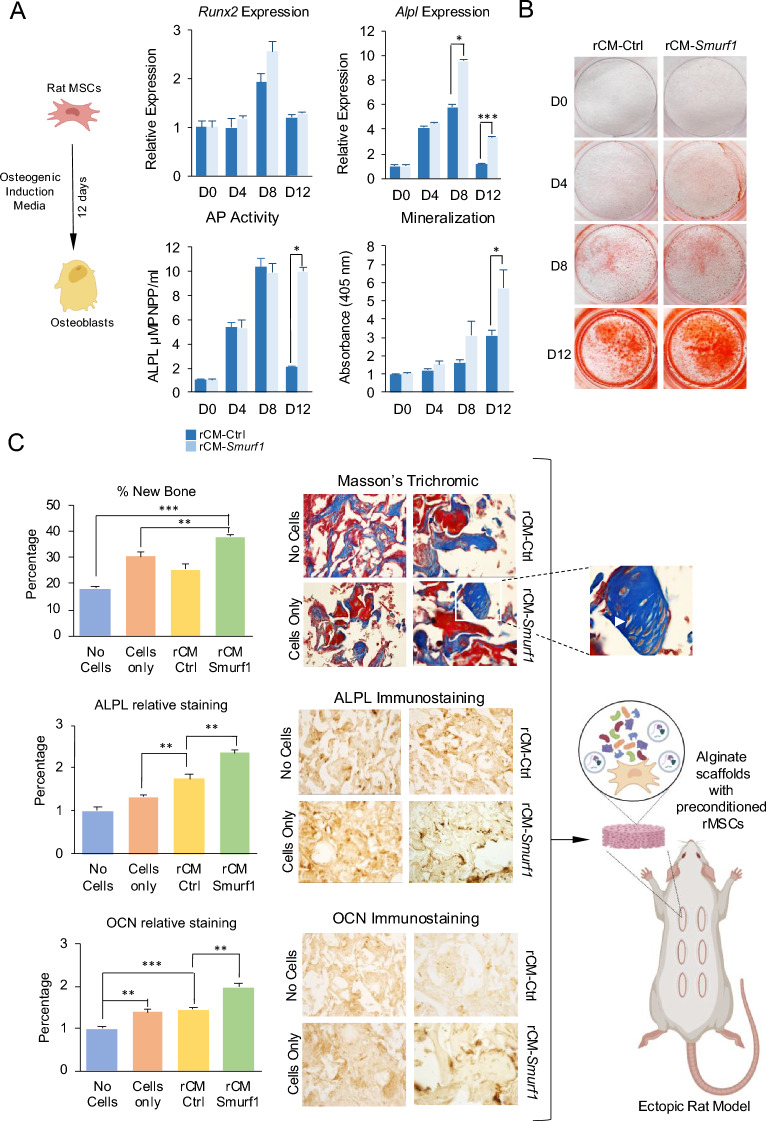
Methods: Conditioned media (CM) from Smurf1-silenced rat (rCM-Smur1) and human MSCs (hCM-Smurf1) was collected and analyzed. Pro-osteogenic potential was assessed by measuring in vitro mineralization in human and rat MSCs cultures. In vivo, studies were conducted using a rat ectopic bone formation model and a post-menopausal osteoporotic mouse model. Additionally, primary human osteoporotic MSCs were preconditioned with hCM-Smurf1, and their osteogenic capacity was compared to that induced by BMP2 treatment. Ex vivo, human bone explants were treated with hCM-Smurf1 to assess anti-resorptive effects. Proteomic analysis of the soluble and vesicular CM fractions identified key proteins involved in bone regeneration.

Results: CM from Smurf1-silenced MSCs significantly enhanced mineralization in vitro and bone formation in vivo. Preconditioning human osteoporotic MSCs with hCM-Smurf1 significantly increases in vitro mineralization, with levels comparable to those achieved with BMP2 treatment. Additionally, in ex vivo human bone cultures, treatment with hCM-Smurf1 significantly reduced RANKL expression without affecting OPG levels, indicating an anti-resorptive effect. In vivo, CM from Smurf1-silenced MSCs significantly increased bone formation in a rat ectopic model, and its local administration reduced trabecular bone loss by 50% in a post-menopausal osteoporotic mouse model after a single administration within just four weeks. Proteomic analysis revealed both soluble and vesicular fractions of hCM-Smurf1 were enriched with proteins essential for ossification and extracellular matrix organization, enhancing osteogenic differentiation.

Conclusions: The Smurf1-silenced MSCs' secretome shows potent osteogenic and anti-resorptive effects, significantly enhancing bone formation and reducing bone loss. This study provides compelling evidence for the therapeutic potential of Smurf1-silenced MSC-derived secretome as a non-toxic and targeted treatment for osteoporosis. These findings warrant further in vivo studies and clinical trials to validate its therapeutic efficacy and safety.

Keywords: Smurf1; Mesenchymal stem cells; Osteoporosis; Secretome.


Conflict of interest statement

Declarations. Ethics approval and consent to participate: The study was conducted according to the guidelines of the Declaration of Helsinki. This study was approved by the Institutional Bioethics Committee of the University of Cantabria (Reference 2015/21; Project Title: Enhancing Mesenchymal Stem Cells Osteogenic Capacity Through Modulation of the Bone Marrow Microenvironment; date of approval 04–11-22) and the Consejería de Agricultura y Ganadería de Cantabria (Reference PI-08–21; Project Title: Modulation of the Regenerative Capacity of Mesenchymal Stem Cells for Their Use in Therapies Applied to the Musculoskeletal System; date of approval 01–11-21). Bone samples were obtained from the patients after anonymization. Informed consent was obtained from all the subjects involved in the study. Consent for publication: Not applicable. Competing interests: The authors declare that they have no competing interests.

Figures

Fig. 1
Fig. 1
In vitro and in vivo analysis of rMSCs-CM pro-osteogenic potential. (A) Top panel. Relative expression levels of osteogenic markers (Runx2 and Alpl) in primary rat MSCs were assessed using semi-quantitative PCR after 48-h of pre-treatment with either rCM-Smurf1 or rCM-Ctrl. Bottom panel. Left graph illustrates the alkaline phosphatase (AP) activity in rMSCs cells preconditioned with rCM-Smurf1 or rCM-Ctrl. Quantification of in vitro mineralization is shown in the bottom panel, right part, of the figure. (B) Alizarin Red staining measuring mineralization was performed every four days during osteogenic differentiation. The images display results from representative samples. For all graphs, results are presented as means ± SEM. (n = 3) with each sample analyzed in technical triplicates. (C) Masson’s trichrome staining of ectodermically implanted scaffolds 8 weeks after implantation. Images show histological analysis of sections obtained from decalcified implants. Collagen of the extracellular bone matrix stained in dark blue. White arrowheads indicate osteocytes‐like cells surrounded by lacunae and immersed in the mineralized matrix (Magnification × 4). A higher magnification shows osteocyte cells surrounded by osteocytic lacunae in CM-Smurf1 (Magnification × 9). Histological sections of the ectodermic scaffolds stained by immunohistochemistry with specific antibody for ALPL and OCN (Magnification × 4). Graphs show the quantification of new bone formation and ALPL and OCN levels observed in the histological sections. Results are presented as means ± SEM. *:p-value < 0.05; **: p-value < 0.01; ***:p-value < 0.001. (n = 3)
Fig. 2
Fig. 2
Pro-osteogenic effect of hCM-SMURF1 on MSCs and bone samples from osteoporotic patients. (A) Top panel. In vitro osteogenic potential of primary human MSCs isolated from the femoral head of osteoporotic patients preconditioned with CM-Ctrl, CM-SMURF1 or BMP2. Normalized results of a quantitative PCR measuring the expression of key osteogenic markers RUNX2, ALPL. Bottom panel left. Graphical representation of the activity of the alkaline phosphatase enzyme. Bottom panel right. Quantification of Alizarin Red staining. Results are presented as means ± SEM (n = 23). *: p-value < 0.05; ***: p -value < 0.001). (B) Alizarin Red staining of representative samples. All analysis were performed after at 20 days of osteogenic differentiation. (C) Gene expression analysis of RANKL, OPG, M-CSF and BGLAP in human osteoporotic bone samples cultured ex vivo in 50% of CM-CTRL or CM-SMURF1 for 4 days measured by qPCR. Results are presented as means ± SEM. *: p-value < 0.05; **: p -value < 0.01; *** p-value < 0.001. (n = 18)
Fig. 3
Fig. 3
Analysis of the hCM-SMURF1 effects on basic cellular functions. (A) Results from the MTT [3-(4,5-dimethylthiazol-2-yl)−2,5-diphenyltetrazolium bromide] proliferation assay performed during 9 days after 48 h of exposition to normal culture media or to the different CMs (hCM-UT = CM from un-transfected cells). The results are presented as ratios of MTT absorbance between consecutive days. *: p-value < 0.05. (n = 3) (B) Wound healing studying the influence of the different CM on cell migration. Quantification of empty area using ImageJ image processor confirmed the absence of significant changes. Graph represents the average area in five different time-points for each of the experimental conditions. Bars represent the SEM. (C) Chemotactic response of MSCs to the different conditioned media was measured using transwell inserts. Media with SDF-1α was used as a positive control. For all experiments, graph represents the average values of five or three experiments, as indicated. Bars show standard deviation of the mean values. *: p-value < 0.05; **: p-value < 0.01; ***: p-value < 0.001
Fig. 4
Fig. 4
Characterization of the vesicular fractions isolated from CM-Ctrl and CM-SMURF1. (A) Nanoparticle Tracking Analysis (NTA) showing particle size distribution of isolated EVs (B) Transmission Electronic Microscopy (TEM) image of EVs isolated from CM-Ctrl and CM-SMURF1. Scale bar 50 nm. (C) Flow cytometry analysis using a bead-bound capture antibody system for the detection extracellular vesicle (EVs) markers. CD63 and CD9. (D) Confocal microscopy images shown EV stained with Vybrant CM Dil (red) and MSCs stained with Phalloidin (green) after two hours of exposition of cells to the stained EVs. Images obtained were composed by ImageJ
Fig. 5
Fig. 5
Pro-osteogenic activity of the soluble (SF) and vesicular (VF) fractions of CM-SMURF1. (A) Top Panel. Expression of osteogenic markers, alkaline phosphatase activity and mineralization at day 16 of osteogenic differentiation after preconditioning with either the soluble (SF) or vesicular (VF) fractions of the hCM-SMURF1. RUNX2 and ALPL expression, as well as alkaline phosphatase activity and mineralization show significant increases in SF of hCM-SMURF1 in relation to hCM-Ctrl. VF do not impact RUNX2, ALPL or alkaline phosphatase activity, however significant enhancement in mineralization is observed. Results are presented as means ± SEM (n = 3). *: p-value < 0.05; **: p-value < 0.01; ***: p-value < 0.001. (B) Alizarin Red staining of representative samples reflecting the stimulation of mineralization in the SF and VF of hCM-SMURF1. (C). Volcano plot showing differently expressed proteins between hCM-Ctrl and hCM-SMURF1 for both SF and VF. Statistical significance was determined using an adjusted t-test against CM-Ctrl, with a p-value threshold of 0.05 and a fold change of 2 or greater. (D) Validation of relative expression by quantitative PCR of 2 genes overexpressed in soluble fraction (SPARC and CCN2) and 2 genes overexpressed in vesicular fraction (PREPL and FMOD) of CM-SMURF1. Results are presented as means ± SEM. *: p-value < 0.05. (n = 5)
Fig. 6
Fig. 6
MicroCT analysis of the osteo-regenerative effect of the local intraosseous administration of the CM-Smurf1 in a post-menopausal osteoporotic mouse model. (A) Experiment outline, (B) representative micro-CT coronal and trans-axial images femurs from Non-Ovariectomized mice (No-OVX), OVX mice treated with NaCl (NaCl), OVX mice treated with the CM-Ctrl (CM-Ctrl) and OVX mice treated with CM-Smurf1 (CM-Smurf1), (C) bone volume/tissue volume (BV/TV; %), trabecular number (Tb.N), trabecular thickness (Tb.Th) and space between trabecula were analyzed by using a Quantum GX micro-CT imaging system (PerkinElmer). Results are presented as means ± SEM (n = 5).*: p-value < 0.05; **: p -value < 0.01

References

    1. Yadav VK, Balaji S, Suresh PS, Liu XS, Lu X, Li Z, Guo XE, Mann JJ, Balapure AK, Gershon MD, Medhamurthy R, Vidal M, Karsenty G, Ducy P. Pharmacological inhibition of gut-derived serotonin synthesis is a potential bone anabolic treatment for osteoporosis. Nat Med. 2010;16(3):308–12. - PMC - PubMed
    1. Khosla S, Riggs BL. Pathophysiology of age-related bone loss and osteoporosis. Endocrinol Metab Clin North Am. 2005;34(4):1015–30. - PubMed
    1. Simpson ER. Sources of estrogen and their importance. J Steroid Biochem Mol Biol. 2003;86(3–5):225–30. - PubMed
    1. Riggs BL, Melton LJ 3rd. The prevention and treatment of osteoporosis. N Engl J Med. 1992;327(9):620–7. - PubMed
    1. Skjodt MK, Frost M, Abrahamsen B. Side effects of drugs for osteoporosis and metastatic bone disease. Br J Clin Pharmacol. 2019;85(6):1063–71. - PMC - PubMed

MeSH terms

LinkOut - more resources