. 2025 Feb 15;39(3):e70373.
doi: 10.1096/fj.202401694R.

Prodomain processing controls BMP-10 bioactivity and targeting to fibrillin-1 in latent conformation

Affiliations

Prodomain processing controls BMP-10 bioactivity and targeting to fibrillin-1 in latent conformation

Chara E S Spanou et al. FASEB J. .

Abstract

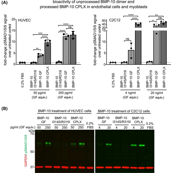
Bone morphogenetic protein 10 (BMP-10) is crucial for endothelial cell signaling via activin receptor-like kinase 1 (ALK1), a pathway central to vascular homeostasis and angiogenesis. Dysregulated BMP-10 signaling contributes to cardiovascular diseases and cancer, highlighting the need to control ALK1-mediated endothelial responses to BMP-10 for therapeutic development. BMP-10 biosynthesis involves processing by proprotein convertases (PPCs) resulting in a non-covalently associated prodomain-growth factor (PD-GF) complex (CPLX), similar to other TGF-β superfamily ligands. However, the molecular requirements for BMP-10 bioactivity remain unclear. We investigated how PPC processing impacts BMP-10 structure, bioactivity, and its interaction with the extracellular matrix (ECM) protein fibrillin-1. Molecular dynamics simulations post-in silico cleavage of the BMP-10 dimer model as well as negative staining and transmission electron microscopy (TEM) revealed that PD processing increases BMP-10 flexibility converting it from a latent wide-angle conformation to a bioactive CPLX which can adopt a V-shape with tighter angle. Only processed BMP-10 demonstrated high potency in HUVEC and C2C12 cells and robust binding to immobilized BMP receptors. Circular dichroism and interaction studies revealed that the N-terminal region of the BMP-10 PD is rich in alpha-helical content, which is essential for efficient complexation with the BMP-10 GF. Binding studies and TEM analyses showed that only the processed BMP-10 CPLX interacts with the N-terminal region of fibrillin-1, causing a conformational change that renders it into a closed ring-shaped conformation. These findings suggest that PD processing induces specific folding events at the PD-GF interface, which is critical for BMP-10 bioactivity and its targeting to the ECM.

Keywords: bone morphogenetic protein; complex; conformational change; electron microscopy; furin; growth factor; molecular dynamics; molecular modeling; proprotein convertases (PPCs); single particle analysis.


Figures

FIGURE 1
FIGURE 1
Generation of unprocessed BMP‐10 dimer. (A) Schematic representation illustrating the BMP‐10 and ‐7 PPC cleavage sites together with the furin cleavage consensus site. (B) Western blot analysis of BMP‐10 PD in HEK293 supernatants transfected with full‐length BMP‐10, or the BMP‐10 I314S/R315I mutant construct. (C) Progressive extracellular processing of the BMP‐10 I314S/R315I mutant after prolonged contact (2–7 days) with the cell layer in serum‐free conditioned medium. (Upper panel) Coomassie‐stained SDS–PAGE gels under reducing conditions after Ni‐NTA affinity chromatography of 500 mL medium collected after 2 days, 7 days, and more than 7 days in contact with the cell layer. (Lower panel) Fraction of processed BMP‐10 as a percentage of the total BMP‐10 signal was determined for each fraction by densitometric analysis of corresponding western blots using the anti‐BMP‐10 PD antibody. (D) (left) Schematic representation illustrating the substitution of the endogenous BMP‐10 furin site with the BMP‐7 PPC site. (right) Coomassie‐stained SDS gels under non‐reducing and reducing conditions after Ni‐NTA affinity chromatography of 500 mL of conditioned medium from HEK293 cells overexpressing the BMP‐10 I314S/R315I mutant.
FIGURE 2
FIGURE 2
Generation and purification of the processed BMP‐10 CPLX. (A) 1. Coomassie‐stained SDS–PAGE gel of elution fractions containing BMP‐10 CPLX from the condensed conditioned medium after HiTrap Q HP anion exchange chromatography, 2. Coomassie‐stained SDS–PAGE gel of elution fractions after HiTrap Q HP anion exchange chromatography of the fraction highlighted in blue in 1. panel, 3. (left) SEC elution profile of processed BMP‐10 CPLX, (right) Coomassie‐stained SDS–PAGE gel after SEC purification of the fraction highlighted in gray in 2. panel, shown under reducing and non‐reducing conditions and native‐PAGE of the SEC purified peak fraction. Panel. (B) (left) Coomassie‐stained SDS–PAGE and native PAGE gels of BMP‐10 PD, the unprocessed BMP‐10 dimer, and the processed BMP‐10 CPLX. (right) Schematic representation illustrating the PD–GF arrangement in processed and unprocessed BMP‐10.
FIGURE 3
FIGURE 3
The processed BMP‐10 CPLX is bioactive towards HUVEC and C2C12 cells while the unprocessed BMP‐10 dimer is latent. (A) Quantification of western blot analysis for HUVEC and C2C12 cells treated for 45 min with the indicated BMP‐10 proteins. pSMAD1/5/9 intensity was normalized to GAPDH intensity, and values were expressed as fold change relative to 0.2% FBS control. Each data point represents the mean value of duplicates, derived from three independent experiments (N = 3). Error bars represent the overall mean ± SD. One‐way ANOVA with multiple comparisons. *p ≤ .05; **p ≤ .01; ***p ≤ .001; ****p ≤ .0001. (B) Representative western blots of cell lysates of HUVEC and C2C12 cells after 45 min stimulation with growth factor equivalents employing anti‐pSMAD1/5/9 and anti‐GAPDH antibodies.
FIGURE 4
FIGURE 4
The processed BMP‐10 CPLX binds robustly to BMP receptors whereas the unprocessed dimer shows no receptor interaction. Sensorgrams of SPR interaction studies of soluble BMP‐10 GF, processed BMP‐10 CPLX, and unprocessed BMP‐10 dimer flowed over immobilized BMP receptors. Soluble analytes were injected onto immobilized BMPRII, ALK‐1, and ENG at concentrations ranging from 0 to 80 nM. K D s were calculated from three independent experiments (N = 3).
FIGURE 5
FIGURE 5
The N‐terminal region of the BMP‐10 PD containing the α1‐helix interacts with the BMP‐10 GF (A) CD spectroscopy analysis of BMP‐9 PD, BMP‐10 PD, and N‐10/C‐9 fusion PD expressed in E. coli. BMP‐10 PD obtained from BMP‐10 CPLX expression in HEK293 cells served as a control together with the in silico computed CD spectra of the BMP‐9 PD based on the atomic model 4YCG. (B) Secondary structure maps of BMP‐9 and BMP‐10 PDs were generated based on the obtained CD data. The position of α‐helices (red) and β‐sheets (blue) was guided by previously reported secondary structure predictions. Glycine and proline residues that are known to prevent the formation of alpha helices are marked in gray. BMP‐9 and BMP‐10 PD sequence alignment was conducted using the MultAlin online tool. (C) Sensorgrams from SPR binding studies of BMP‐10 GF flowed over immobilized BMP‐9 PD, BMP‐10 PD, and the N‐10/ C‐9 fusion PD at concentrations from 0 to 80 nM (representative sensograms of three independent experiments are shown).
FIGURE 6
FIGURE 6
BMP‐10 CPLX assumes a V‐shape conformation while unprocessed BMP‐10 shows a conformation with a wider angle. (A) Single particle transmission EM class averages of the unprocessed BMP‐10 dimer from negatively stained images. (B) EM envelope of the 3D reconstruction of the unprocessed BMP‐10 dimer. (C) Negative staining TEM images of unprocessed and processed BMP‐10 at a 1:1 molar ratio, revealing BMP‐10 molecules with both V‐shape as well as with wide‐angle conformations. (D) Quantification based on 600 BMP‐10 CPLX particles per field across 61 different fields, showed an equal presence of molecules with a V‐shape and wide angle (1:1 molar ratio). Scale bar: 20 nm.
FIGURE 7
FIGURE 7
Models of unprocessed BMP‐10 dimer and processed BMP‐10 CPLX. (A) Model of the unprocessed BMP‐10 dimer showing the α1‐helix (marked in blue) masking ALK‐1‐binding residues Y358, P359, and I371 on the GF (marked in red). (B) Model of the processed BMP‐10 CPLX where the same GF residues (marked in cyan) are accessible for ALK‐1 receptor engagement. Processing reduces the distance between PD arms by about 20 Å as indicated. PPC cleavage sites are marked in light blue.
FIGURE 8
FIGURE 8
Prodomain processing alters BMP‐10 surface charge. (A) (left) Coomassie R‐stained native PAGE gel of unprocessed BMP‐10 I314S/R315I and processed BMP‐10 CPLX indicates different running behavior between the two BMP‐10 processing variants. (right) Coomassie‐stained SDS‐PAGE gels of unprocessed BMP‐10 I314S/R315I and processed BMP‐10 CPLX were analyzed under reducing and non‐reducing conditions. (B) Experimental and theoretical CD spectra of BMP‐10 PD, GF, unprocessed BMP‐10 I314S/R315I, and processed CPLX. (C) Electrostatic surface coloring of unprocessed BMP‐10 or processed BMP‐10 CPLX models using ChimeraX.
FIGURE 9
FIGURE 9
BMP‐10 is targeted to fibrillin‐1. (A) Co‐localization of BMP‐10 PD with deposited fibrillin‐1 fibers was detected in primary murine aortic VSMC culture. (B) SPR interaction experiment shows that only processed BMP‐10 interacts with the immobilized N‐terminal region of fibrillin‐1.
FIGURE 10
FIGURE 10
Processed BMP‐10 CPLX interacts with fibrillin‐1 and is rendered into a closed ring‐shape conformation. (A) TEM analysis after incubation of the N‐terminal fibrillin‐1N‐terminal region with a 1:1 mixture of processed and unprocessed BMP‐10. (top, left) Representative negative staining TEM images showing the globular shape of the N‐terminal region of fibrillin‐1 (start_EGF4). (top, right; bottom left) TEM analysis of unbound BMP‐10 molecules. Scale bars: 30 nm (overview) or 20 nm (magnified micrographs). (bottom, right) Quantification of unbound BMP‐10 particles showing a significant increase of molecules with wide angle (80%) versus tight angle (20%). (B) TEM analysis reveals the presence of ring‐shaped BMP‐10 molecules upon the addition of fibrillin‐1 (start_EGF4). The apparent position of globular N‐terminal fibrilin‐1 molecules is indicated by white arrow heads in one representative example. Scale bar: 20 nm. (C) Model of the closed‐ring BMP‐10 CPLX. In this model, the type II receptor binding site: 405GVVTYKF411 on BMP‐10 GF (light green) is masked by the α2‐helix, while ALK‐1 binding residues on the GF remain accessible. The BMP‐10 binding sites within FUN are labeled in black and yellow, and the fibrillin‐1 binding site in BMP‐10 PD is labeled in orange. PD residues are labeled in purple and GF residues in dark green.
FIGURE 11
FIGURE 11
Working model describing targeting, sequestration, and activation of BMP‐10 depending on its processing status. BMP‐10 is produced systemically by hepatic stellate cells and locally by cardiovascular resident cells. Its activation depends on the localization of PPCs. When PPCs are present intracellularly, BMP‐10 is first processed and secreted in a bioactive form, which is then sequestered by fibrillin microfibrils in latent pools. From these pools, BMP‐10 can be reactivated locally through PD‐proteolytic cleavage, releasing the bioactive GF. Conversely, when PPCs are present extracellularly, BMP‐10 is secreted in an unprocessed, latent form. Activation occurs via PPC‐mediated cleavage of the covalent bond between the PD and the GF, which increases flexibility in the α1 helix, enabling specific GF residues to interact with ALK‐1 and initiate long‐range BMP signaling. These distinct activation pathways mediate either short‐range or long‐range signaling, activating unique signaling cascades during development and homeostasis.

References

    1. Shi Y, Massague J. Mechanisms of TGF‐beta signaling from cell membrane to the nucleus. Cell. 2003;113:685‐700. - PubMed
    1. Gregory KE, Ono RN, Charbonneau NL, et al. The prodomain of BMP‐7 targets the BMP‐7 complex to the extracellular matrix. J Biol Chem. 2005;280:27970‐27980. - PubMed
    1. Brown MA, Zhao Q, Baker KA, et al. Crystal structure of BMP‐9 and functional interactions with pro‐region and receptors. J Biol Chem. 2005;280:25111‐25118. - PubMed
    1. Ge G, Hopkins DR, Ho WB, Greenspan DS. GDF11 forms a bone morphogenetic protein 1‐activated latent complex that can modulate nerve growth factor‐induced differentiation of PC12 cells. Mol Cell Biol. 2005;25:5846‐5858. - PMC - PubMed
    1. Jiang H, Salmon RM, Upton PD, et al. The prodomain‐bound form of bone morphogenetic protein 10 is biologically active on endothelial cells. J Biol Chem. 2016;291:2954‐2966. - PMC - PubMed

LinkOut - more resources