Review
. 2025 Feb 18;14(4):e036952.
doi: 10.1161/JAHA.124.036952. Epub 2025 Feb 8.

Skeletal Muscle Pathology in Pulmonary Arterial Hypertension and Its Contribution to Exercise Intolerance

Affiliations
Review

Skeletal Muscle Pathology in Pulmonary Arterial Hypertension and Its Contribution to Exercise Intolerance

Thaís C F Menezes et al. J Am Heart Assoc. .

Abstract

Pulmonary arterial hypertension is a disease of the pulmonary vasculature, resulting in elevated pressure in the pulmonary arteries and disrupting the physiological coordination between the right heart and the pulmonary circulation. Exercise intolerance is one of the primary symptons of pulmonary arterial hypertension, significantly impacting the quality of life. The pathophysiology of exercise intolerance in pulmonary arterial hypertension is complex and likely multifactorial. Although the significance of right ventricle impairment and perfusion/ventilation mismatch is widely acknowledged, recent studies suggest pathophysiology of the skeletal muscle contributes to reduced exercise capacity in pulmonary arterial hypertension, a concept explored herein.

Keywords: exercise intolerance; mitochondrial dysfunction; oxygen pathway; pulmonary hypertension; skeletal muscle dysfunction.


Conflict of interest statement

None.

Figures

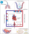
Figure 1
Figure 1. Exercise intolerance in patients with pulmonary arterial hypertension.
RV indicates right ventricle; SKM, skeletal muscle; V/Q, relation between ventilation and perfusion; and VD/VT, dead space ventilation ratio.
Figure 2
Figure 2. Illustration of the 6 steps of O2 pathway from environment to mitochondria.
O2 indicates oxygen.
Figure 3
Figure 3. The relationship between uptake VO2 and microvascular oxygen partial pressure.
Fick's law (blue line) describes the diffusion of oxygen from capillaries to mitochondria. Fick's principle (black line) shows VO2 as the product of oxygen supply and muscle extraction capacity (difference in oxygen content between arterial and venous blood [C(a‐v)O₂]). The point where the 2 curves intersect marks the maximum oxygen consumption (VO₂MAX), which is the maximum VO2 during exercise. C(a‐v)O2 indicates difference in oxygen content between arterial and venous blood; CO, cardiac output; D, distance; O2, oxygen; PO2, oxygen partial pressure; and VO₂MAX, maximum oxygen consumption.
Figure 4
Figure 4. Vascular and skeletal muscle signaling pathways.
A, Skeletal muscle endothelial cells: miR‐126 suppresses PIK3R2 and SPRED1, which negatively regulates VEGF signaling via the PI3 kinase and RAF1 kinase pathways, ultimately affecting angiogenesis. B, Summary schema of protein synthesis and degradation in muscle cells. AKT signaling can increase both the activity of protein synthesis pathways and inhibit protein degradation pathways. TAK1 signaling can increase protein degradation. AKT indicates protein kinase B; ERK, extracellular signal‐regulated kinase; FOXO, Forkhead box O transcription factors; IGF‐1, insulin‐like growth factor 1; miR‐126, microRNA 126; mTOR, mammalian target of rapamycin; MuRF‐1, muscle RING‐finger protein‐1; p38MAPK, p38 mitogen‐activated protein kinase; p70S6, ribosomal protein S6 kinase beta‐1; PI3 kinase, phosphoinositide 3‐kinase; PIK3R2, phosphoinositide‐3‐kinase regulatory subunit 2; RAF1, RAF proto‐oncogene serine/threonine‐protein kinase; SPRED1, sprouty‐related EVH1 domain‐containing protein 1; TAK1, transforming growth factor‐beta‐activated kinase 1; TGF‐β, transforming growth factor‐β; and VEGF, vascular endothelial growth factor.

References

    1. Humbert M, Kovacs G, Hoeper MM, Badagliacca R, Berger RMF, Brida M, Carlsen J, Coats AJS, Escribano‐Subias P, Ferrari P, et al. 2022 ESC/ERS guidelines for the diagnosis and treatment of pulmonary hypertension. Eur Heart J. 2022;43:3618–3731. doi: 10.1093/eurheartj/ehac237 - DOI - PubMed
    1. Waxman AB. Exercise physiology and pulmonary arterial hypertension. Prog Cardiovasc Dis. 2012;55:172–179. doi: 10.1016/j.pcad.2012.07.003 - DOI - PubMed
    1. Batt J, Ahmed SS, Correa J, Bain A, Granton J. Skeletal muscle dysfunction in idiopathic pulmonary arterial hypertension. Am J Respir Cell Mol Biol. 2014;50:74–86. doi: 10.1165/rcmb.2012-0506OC - DOI - PubMed
    1. McLaughlin VV, Shillington A, Rich S. Survival in primary pulmonary hypertension. Circulation. 2002;106:1477–1482. - PubMed
    1. Sun X‐G, Hansen JE, Oudiz RJ, Wasserman K. Exercise pathophysiology in patients with primary pulmonary hypertension. Circulation. 2001;104:429–435. doi: 10.1161/hc2901.093198 - DOI - PubMed

MeSH terms

LinkOut - more resources