. 2025 Feb 8;149(1):16.
doi: 10.1007/s00401-025-02854-x.

Perivascular glial reactivity is a feature of phosphorylated tau lesions in chronic traumatic encephalopathy

Affiliations

Perivascular glial reactivity is a feature of phosphorylated tau lesions in chronic traumatic encephalopathy

Chelsie Osterman et al. Acta Neuropathol. .

Abstract

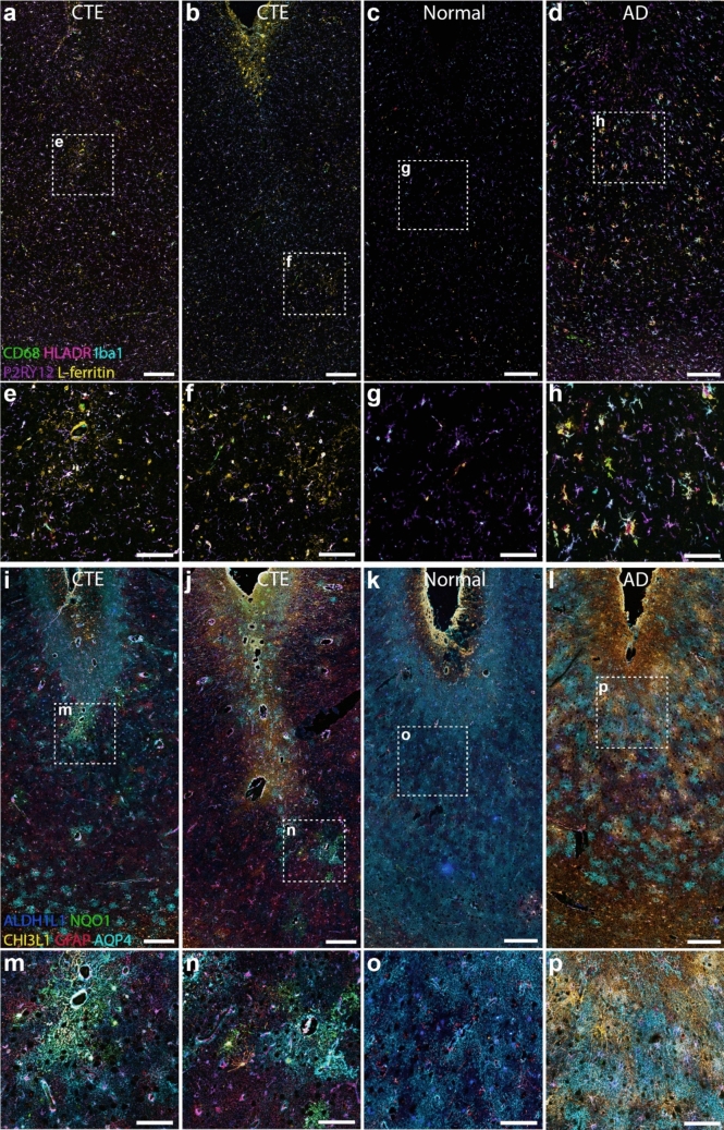
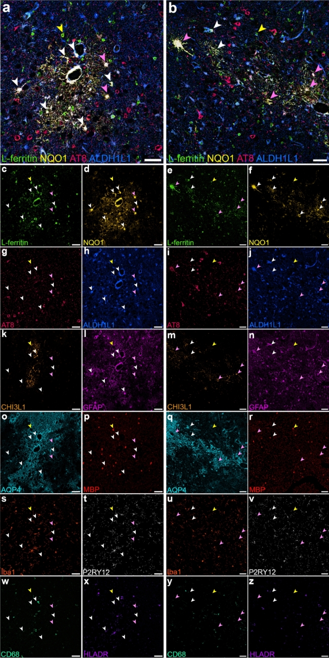
Chronic traumatic encephalopathy (CTE), a neurodegenerative disease associated with repetitive head injuries, is characterised by perivascular hyperphosphorylated tau (p-tau) accumulations within the depths of cortical sulci. Although the majority of CTE literature focuses on p-tau pathology, other pathological features such as glial reactivity, vascular damage, and axonal damage are relatively unexplored. In this study, we aimed to characterise these other pathological features, specifically in CTE p-tau lesion areas, to better understand the microenvironment surrounding the lesion. We utilised multiplex immunohistochemistry to investigate the distribution of 32 different markers of cytoarchitecture and pathology that are relevant to both traumatic brain injury and neurodegeneration. We qualitatively assessed the multiplex images and measured the percentage area of labelling for each marker in the lesion and non-lesion areas of CTE cases. We identified perivascular glial reactivity as a prominent feature of CTE p-tau lesions, largely driven by increases in astrocyte reactivity compared to non-lesion areas. Furthermore, we identified astrocytes labelled for both NAD(P)H quinone dehydrogenase 1 (NQO1) and L-ferritin, indicating that lesion-associated glial reactivity may be a compensatory response to iron-induced oxidative stress. Our findings demonstrate that perivascular inflammation is a consistent feature of the CTE pathognomonic lesion and may contribute to the pathogenesis of brain injury-related neurodegeneration.

Keywords: Astrocyte; Brain injury; Chronic traumatic encephalopathy; Neurodegeneration; Neuroinflammation; Neuropathology; Tau.


Conflict of interest statement

Declarations. Conflict of interest: The authors have no competing interests to declare that are relevant to the content of this article.

Figures

Fig. 1
Fig. 1
Overview of multiplex labelling for tau and neuronal markers in the frontal cortex of CTE cases. Overview of the area at the depth of the cortical sulcus containing a p-tau lesion in two CTE cases (AU2 and AU7, both high-stage CTE) from the Australia Sports Brain Bank (a–b, e–f), and the corresponding area in a representative neurologically normal (c, g) and AD case (d, h). Multiplex labelling for tau pathology markers 4R, 3R, p-tau202-205 (AT8), p-tau231 (T231), MN423 and beta-amyloid are presented (a-d), alongside a higher magnification of the lesion vessel and comparable regions in the AD and normal cases indicated by the dotted box (e–h). Neurofibrillary tangles labelled for 4R, 3R, AT8, and T231 within the perivascular p-tau lesion in CTE cases (a-b, e–f). The AD case also showed extensive neurofibrillary tangles and fibres that were labelled for 4R, 3R, AT8, and T231 (d, h). Tau labelling was absent in the neurologically normal case (c, g). Multiplex labelling for neuronal subtype markers NeuN, calbindin, calretinin, and parvalbumin are presented with AT8 (i-l), along with higher magnification of the regions indicated by the dotted box (m-p). The perivascular tau-positive cells were predominantly located in layer II of the cortex and co-labelled with the neuronal marker NeuN, but not with the interneuron markers calbindin, calretinin, or parvalbumin. Scale bars: 250 µm (a-d, i–l); 100 µm, (e–h, m–p)
Fig. 2
Fig. 2
Overview of multiplex labelling for markers of reactive glia in the frontal cortex of CTE cases. Multiplex labelling of reactive microglia (a–h) and astrocytes (i-p) within the depth of the cortical sulcus containing a p-tau lesion in two CTE cases from the Australia Sports Brain Bank (a–b, e–f) and the corresponding area in a representative normal (c, g) and AD case (d, h). Reactive microglial markers CD68, HLA-DR and L-ferritin, and homeostatic microglial markers Iba1 and P2RY12 are presented (a–d) alongside higher magnification of the lesion vessel and comparable regions in AD and normal cases indicated by the dotted box (e–h). CTE cases show elevated immunoreactivity for L-ferritin but comparable labelling for all other microglial markers compared to normal cases. Reactive astrocyte markers NQO1, CHI3L1, and GFAP, homeostatic astrocyte marker ALDH1L1, and astrocyte water channel marker AQP4 are presented (i-l) alongside higher magnification of the lesion vessel and comparable regions indicated by the dotted box (m–p). Immunoreactivity for reactive astrocyte markers was increased around the lesion vessels in CTE cases, compared to normal cases and the more ubiquitous distribution seen in AD cases. Scale bars: 250 µm (a–d, i–l); 100 µm, (e–h, m–p)
Fig. 3
Fig. 3
RNA and protein distribution for reactive gliosis markers in CTE lesion sulcus. a Spatial gene expression heatmaps generated from the Visium spatial transcriptomics data illustrates the mRNA distribution for NQO1, CHI3L1, GFAP, AQP4 and FTL (L-ferritin) within a lesion sulcus for CTE case AU6 from the Australia Sports Brain Bank. b Immunohistochemistry was performed for the corresponding protein in (a) on a different lesion sulcus from the same case. The p-tau (AT8) labelling for the same tissue sections is shown at the bottom (c–d). Scale bar: 200 µm. In all three cases, focal increases in NQO1, CHI3L1, GFAP, AQP4 and FTL (L-ferritin) mRNA and protein expression are seen in cortical layer 1, the white matter and within the lesion area (yellow arrow) where AT8 tau is highly concentrated. The sulcal border is marked with an asterisk
Fig. 4
Fig. 4
Differentially expressed markers in CTE lesion areas relative to non-lesion areas. a The area of labelling for each marker was measured within a 1mm2 region around the p-tau-positive lesion vessel in the sulci and p-tau-negative non-lesion vessels in the adjacent gyrus. Scale bar 1 mm. b Volcano plot illustrating differentially expressed markers between the lesion and non-lesion areas. Paired t tests (parametric) or Wilcoxon tests (non-parametric) were conducted to assess the statistical significance of the difference in the area of positive labelling between lesion and non-lesion regions of interest. Vertical dashed lines indicate the log2 (Fold change) cut-off of 0.5 (twofold change). Horizontal dashed lines indicate − log10 (p value) cut-off at 0.05. Each red dot represents one marker, with the mean log2 fold change between lesion and non-lesion regions across all CTE cases plotted. The labelled red dots are lesion-associated differentially expressed markers. c An example of the Sholl analysis. Concentric rings radiated from the vessel lumen with 20 µm spacing. Scale bar 50 µm (d–e) Representative images of the labelling for differentially expressed glial reactivity markers in lesion (d) and non-lesion (e) regions from two CTE cases (AU1, BU6) provided by different brain banks. Scale bars: 50 µm. f–k Graphs of the Sholl analysis for lesion and non-lesion vessels, where the area of labelling for each marker was measured within concentric rings up to 300 µm from the vessel lumen. The area of labelling was normalised to the area of the ring, and the values were normalised to the maximum value of the dataset for that case. The mean ± SEM normalised area of labelling for each marker was plotted against the distance from the vessel
Fig. 5
Fig. 5
NQO1 and L-ferritin co-labelling in astrocytes within the CTE lesion. Images of the p-tau lesion from CTE case AU2 (a) and AU1 (b) illustrating co-labelling for glial reactivity markers L-ferritin (c, e) and NQO1 (d, f), with AT8 tau (g, i, pink arrows) and the homeostatic astrocyte marker ALDH1L1 (h, j, white and pink arrows) and L-ferritin+ NQO1+ cells also co-labelled with GFAP (l, n), but not CHI3L1 (k, m) or AQP4 (o, q). A subset of L-ferritin cells also co-labelled with myelin basic protein (p, r, yellow arrows) and a different subset co-labelled for the microglial markers Iba1 (s, u), P2RY12 (t, v), CD68 (w, y), and HLA-DR (x, z). Scale bars 50 µm

References

    1. Bieniek KF, Cairns NJ, Crary JF, Dickson DW, Folkerth RD, Keene CD et al (2021) The second NINDS/NIBIB consensus meeting to define neuropathological criteria for the diagnosis of chronic traumatic encephalopathy. J Neuropathol Exp Neurol 80(3):210 - PMC - PubMed
    1. McKee AC, Cairns NJ, Dickson DW, Folkerth RD, Dirk Keene C, Litvan I et al (2016) The first NINDS/NIBIB consensus meeting to define neuropathological criteria for the diagnosis of chronic traumatic encephalopathy. Acta Neuropathol 131(1):75 - PMC - PubMed
    1. Nowinski CJ, Bureau SC, Buckland ME, Curtis MA, Daneshvar DH, Faull RLM et al (2022) Applying the Bradford Hill criteria for causation to repetitive head impacts and chronic traumatic encephalopathy. Front Neurol 22:13 - PMC - PubMed
    1. Cherry JD, Kim SH, Stein TD, Pothast MJ, Nicks R, Meng G et al (2020) Evolution of neuronal and glial tau isoforms in chronic traumatic encephalopathy. Brain Pathol 30(5):913–925 - PMC - PubMed
    1. Hamlin D, Ryall C, Turner C, Faull RLM, Murray HC, Curtis MA (2024) Characterization of neurofibrillary tangle immunophenotype signatures to classify tangle maturity in Alzheimer’s disease. Alzheimers Dement 20(7):4803 - PMC - PubMed

Publication types

LinkOut - more resources