. 2025 Mar 20;188(6):1711-1728.e21.
doi: 10.1016/j.cell.2024.12.032. Epub 2025 Feb 7.

Molecular basis of convergent evolution of ACE2 receptor utilization among HKU5 coronaviruses

Affiliations

Molecular basis of convergent evolution of ACE2 receptor utilization among HKU5 coronaviruses

Young-Jun Park et al. Cell. .

Abstract

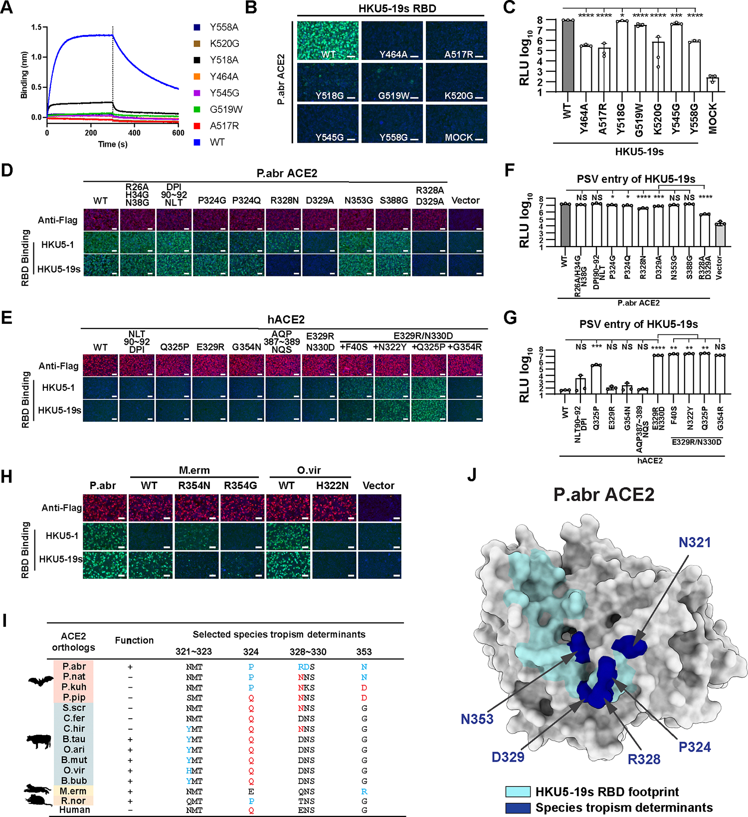
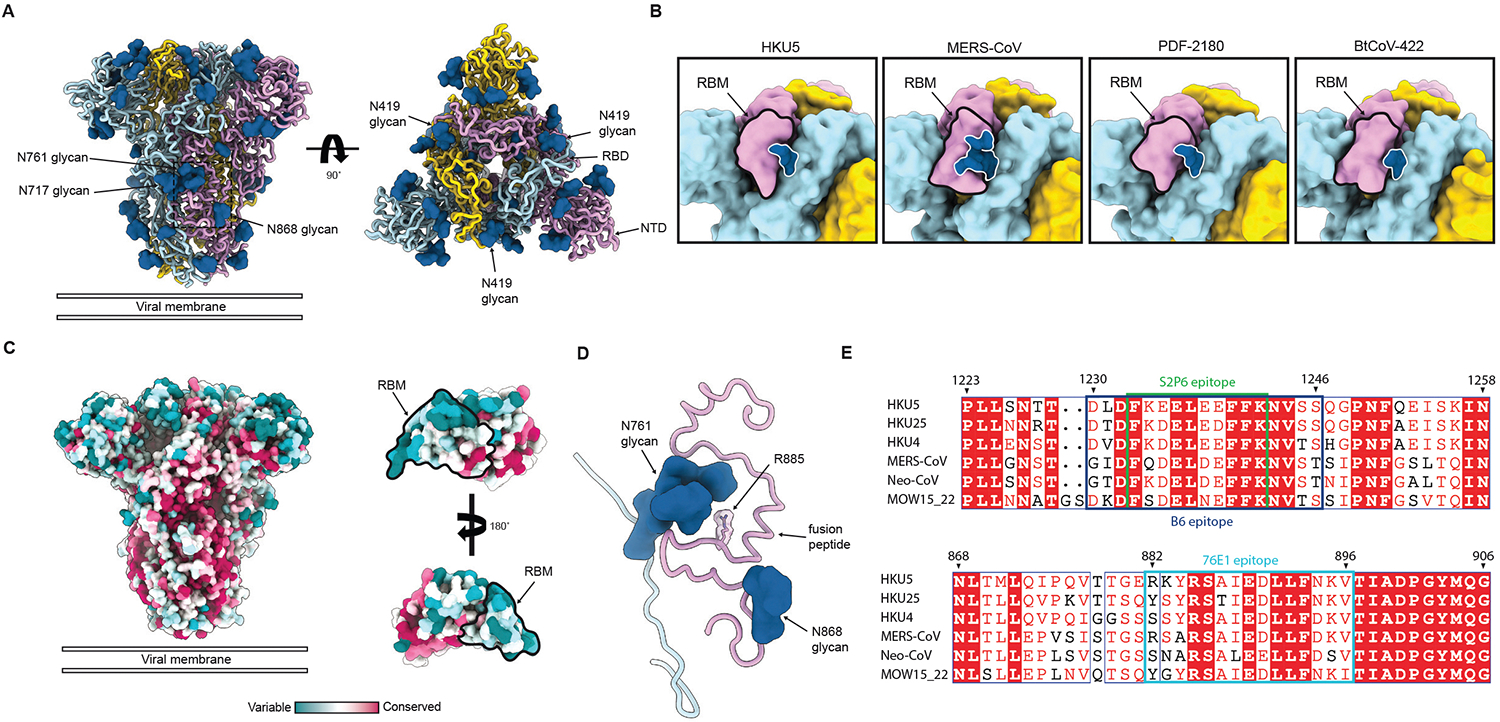
DPP4 was considered a canonical receptor for merbecoviruses until the recent discovery of African bat-borne MERS-related coronaviruses using ACE2. The extent and diversity of ACE2 utilization among merbecoviruses and their receptor species tropism remain unknown. Here, we reveal that HKU5 enters host cells utilizing Pipistrellus abramus (P.abr) and several non-bat mammalian ACE2s through a binding mode distinct from that of any other known ACE2-using coronaviruses. We defined the molecular determinants of receptor species tropism and identified a single amino acid mutation enabling HKU5 to utilize human ACE2, providing proof of principle for machine-learning-assisted outbreak preparedness. We show that MERS-CoV and HKU5 have markedly distinct antigenicity and identified several HKU5 inhibitors, including two clinical compounds. Our findings profoundly alter our understanding of coronavirus evolution, as several merbecovirus clades independently evolved ACE2 utilization, and pave the way for developing countermeasures against viruses poised for human emergence.

Keywords: HKU5; MERS-CoV; antibodies; coronaviruses; merbecoviruses; spillover; viral receptor.


Conflict of interest statement

Declaration of interests H.Y. has submitted a patent application to the China National Intellectual Property Administration for the utilization of propagation-competent VSV to evaluate amplification of ACE2-using merbecoviruses in bat ACE2-expressing cells.

Figures

Figure 1.
Figure 1.. Identification of several mammalian ACE2s as functional HKU5 entry receptors.
(A) Merbecovirus RBD phylogenetic tree based on amino acid sequences defining 6 clades. c/hMERS: camel/human MERS-CoV isolates. Each merbecovirus is listed along with its GenBank ID. The animal symbols represent the hosts in which viruses have been detected. (B) Phylogenetic trees of bat (top) or non-bat (bottom) mammalian ACE2 orthologs based on amino acid sequences, with genera and orders indicated for the bat and non-bat mammalian species, respectively. (C-D) Binding of the HKU5–19s RBD-hFc to and entry of HKU5–19s S VSV pseudovirus (pretreated with 100 μg/ml TPCK-treated trypsin) into HEK293T cells transiently transfected with the indicated bat (C) or non-bat (D) mammalian ACE2 orthologs. Abbr: abbreviations used for species names. Mean values are shown in C and D with n=3 biological replicates. (E-F) Propagation of pcVSV-HKU5–1 (E, left), pcVSV-HKU5–19s (E, right), and wildtype authentic HKU5–1 (F) in human Caco-2 cells (endogenously expressing hACE2) or Caco-2 cells with stable expression of either hACE2 or P.abr ACE2. The propagation of pcVSV-HKU5–1 was detected by the expression of the GFP reporter gene at the indicated hours post-infection (hpi) whereas authentic HKU5–1 was detected by immunofluorescence with an anti-HKU5 N antibody at 24 hpi. The trypsin concentration used is indicated. Exogenous ACE2 expression was confirmed by immunofluorescence using C-terminally-fused FLAG tags. Scale bars: 200 μm. See also Fig S1–S3.
Figure 2.
Figure 2.. Molecular basis of HKU5 recognition of the P. abramus ACE2 receptor.
(A) Ribbon diagrams in two orthogonal orientations of the cryo-EM structure of the HKU5–19s RBD (light blue) bound to the P. abramus ACE2 peptidase domain (green). (B-C) Zoomed-in views of the interface highlighting key interactions between the HKU5–19s RBD and P.abr ACE2. Selected polar interactions are shown as black dotted lines. (D) Polymorphism of ACE2-interacting residues (receptor-binding motif, RBM) among HKU5 isolates (Genbank ID: HKU5–17s: AGP04930.1, HKU5–24s: AGP04936.1, HKU5–33s: AGP04943.1, HKU5–27s: AGP04938.1, HKU5–20s: AGP04933.1, HKU5–28s: AGP04939.1, HKU5–32s: AGP04942.1, HKU5–21s: AGP04934.1, HKU5–19s: AGP04932.1, HKU5–1-LMH03F: NC_009020.1, HKU5–31s: AGP04941.1, HKU5–22s: AGP04935.1, HKU5–30s: AGP04940.1, HKU5–2: ABN10884.1, HKU5–1: YP_001039962.1, HKU5-JX2020-Q257: WCC63324.1, HKU5-JX2020-Q279: WCC63520.1, HKU5-GD2017-Q262: WCC63369.1, HKU5r-BY140535: AWH65910.1) shown as logoplot. Amino acids are colored according to their chemical properties: hydrophobic (black), polar non-charged (green), positively charged (blue) and negatively charged (red). (E) Amino acid sequence alignment of the HKU5–19s, HKU5–33s and HKU5–1 RBMs. Residues indicated with a red background are strictly conserved whereas residues indicated with a white background show variability. The residue numbering corresponds to HKU5–19s. HKU5–33s insertions are shown as black dots. The blue lines indicate residues outside the RBM shown for visualization purposes around the HKU5–33s insertions. (F-H) Biolayer interferometry analysis of the P.abr ACE2 ectodomain binding to the HKU5–19s (F), HKU5–33s (G) or HKU5–1 (H) RBDs immobilized on streptavidin (SA) biosensors. Binding avidities were determined by steady state kinetics and are reported as apparent affinities (KD,app). The vertical dotted lines indicate the transition between association and dissociation phases. One representative out of two biological replicates with independently produced batches of proteins are shown for F-H. (I-L) Comparison of the structures of the P.abr ACE2-bound HKU5 RBD (PDB 9D32, this study), the P.pip ACE2-bound NeoCoV RBD (PDB 7WPO), hDPP4-bound MERS-CoV RBD (PDB 4KR0) and hDPP4-bound HKU4 RBD (PDB 4QZV). See also Fig S4–S5, S8 and Tables 1–2.
Figure 3.
Figure 3.. HKU5 molecular determinants of ACE2 host species tropism.
(A) Binding of the P.abr ACE2 construct comprising the peptidase and the dimerization domains (residues 20–724) at a concentration of 100 nM to the wildtype (WT) HKU5–19s and to the listed RBD interface mutants immobilized on biolayer interferometry streptavidin (SA) biosensors. (B-C) RBD-hFc binding (B) and pseudovirus entry (C) efficiencies of HKU5–19s mutants in HEK293T cells transiently expressing P.abr ACE2. (D-E) HKU5–19s and HKU5–1 RBD-hFc binding to HEK293T cells transiently expressing the indicated P.abr ACE2 or hACE2 mutants assessed by immunofluorescence. (F-G) HKU5–19s S VSV pseudovirus entry into HEK293T cells transiently expressing the indicated P.abr ACE2 or hACE2 mutants. (H) HKU5–19s and HKU5–1 RBD-hFc binding to HEK293T cells transiently expressing wildtype and mutated M.erm ACE2 or O.vir ACE2. (I) Summary of ACE2 residues governing species tropism for HKU5–19s. Favorable and unfavorable residues in ACE2 orthologs are highlighted in blue and red, respectively, using P.abr ACE2 residue numbering. (+): functional receptor; (−): non-functional receptor. (J) Critical determinants of HKU5–19s receptor species tropism are mapped in navy color on the P.abr ACE2 structure rendered as a gray surface. The rest of the HKU5–19s RBD footprint is shown in cyan. Data are shown as the MEAN ± SD of 3 biological replicates for C, F, and G. Statistical analyses used unpaired two-tailed t-tests: *: p < 0.05,**: p < 0.01, ***: p < 0.005, and ****: p < 0.001, NS: not-significant. One representative out of two biological replicates with independently produced batches of proteins are shown for A. Data representative of two independent experiments for C, F and G each performed with three biological replicates. Scale bars: 100 μm.
Figure 4.
Figure 4.. HKU5 utilization of artiodactyl and human ACE2 receptors
(A) Ribbon diagrams in two orthogonal orientations of the cryo-EM structure of the HKU5–19s RBD (light blue) bound to the B.tau ACE2 peptidase domain (light green). (B-C) Zoomed-in views of the interface highlighting key interactions between the HKU5–19s RBD and B.tau ACE2. Selected polar interactions are shown as black dotted lines. (D) AlphaFold3-predicted structure of the interface between the HKU5–19s Y560K RBD and hACE2. (E-G) Biolayer interferometry analysis of the B.tau ACE2 ectodomain binding to the HKU5–19s (E), HKU5–1 (F) and HKU5–33s (G) RBDs. Binding avidities were determined by steady state kinetics and are reported as apparent affinities (KD,app). (H) Evaluation of binding of the hACE2 ectodomain dimer to the immobilized wildtype HKU5–19s and HKU5–1 RBDs and to the HKU5–19s Y560K mutant RBD. In panels E-H, the vertical dotted lines indicate the transition between association and dissociation phases. One representative out of two biological replicates with independently produced batches of proteins are shown for E-H. (I-J) Pseudovirus entry efficiencies in HEK293T cells transiently expressing hACE2 (I) or P.abr ACE2 (J) of wildtype and Y560K HKU5–19s S VSV and wildtype HKU5–33s S VSV. Pseudoviruses were either used untreated or after incubation with 100 μg/mL trypsin for 15 min at room temperature before addition of trypsin soybean inhibitor to stop the reaction. Mock: VSV pseudovirus produced without transfecting an S construct. Each bar represents the geometric mean of three biological replicates with geometric SD and each dot represents the geometric mean of three technical replicates. See also Fig S3–S5, S8 and Table 1.
Figure 5.
Figure 5.. Architecture and conservation of the HKU5 S glycoprotein trimer.
(A) Ribbon diagrams in two orthogonal orientations of the cryo-EM structure of prefusion HKU5–19s S. The dotted black box outlines the S2’ cleavage site and fusion peptide region shown in (D). (B) Close-up view of the receptor-binding motif (RBM, outlined in black) and shielding glycan (outlined in white) in HKU5–19s, MERS-CoV (PDB 6Q04), PDF-2180 (PDB 7U6R), and BtCoV-422 (PDB 8SAK) prefusion S trimers. (C) Conservation analysis using Consurf of S residues across the merbecovirus phylogeny shown in Fig. 1A mapped on prefusion HKU5–19s S and the RBD. Residue conservation ranges from variable (teal), average (white), to conserved (red). (D) Zoomed-in view of the HKU5 S2’ cleavage site (including R885) and adjacent fusion peptide region highlighting the glycans at position N761 and N868 limiting access. (E) Amino acid sequence alignment of one representative S glycoprotein from each of the six merbecovirus clades focused on the epitopes recognized by the S2P6 (green) and B6 (navy) stem helix-directed antibodies as well as the 76E1 (cyan) fusion peptide-directed antibodies. Residues indicated with a red background are strictly conserved whereas residues indicated with a white background show variability. The residue numbering corresponds to HKU5–19s. Alignment and visualization were done using ESPript. See also Fig S6 and Table 1.
Figure 6.
Figure 6.. Identification of countermeasures against HKU5 merbecoviruses.
(A) Neutralization of HKU5–19s S VSV pseudovirus mediated by a panel of MERS-CoV or SARS-CoV-2 infection-elicited polyclonal antibody-containing human plasma. Bars represent the mean of three biological replicates with SD and each data point corresponds to the mean of two technical replicates within each biological replicate carried out with distinct batches of pseudoviruses. Statistical significance was measured using the one sample t test against the maximum VSV entry value of 100%. ns:P > 0.05, *:P≤0.05, **:P≤ 0.01, ***:P≤ 0.001, ****:P≤ 0.0001. (B-C) Evaluation of inhibition of wildtype authentic HKU5–1 propagation in Caco-2 cells stably expressing P.abr ACE2 by the indicated concentration of broadly neutralizing antibodies (B) or small molecule/peptide inhibitors (C). HKU5–1 infection was detected by immunofluorescence using an anti-HKU5 N protein antibody at 24 hpi. Scale bars: 100 μm. See also Fig S7 and Table 3.
Figure 7.
Figure 7.. Coronaviruses have evolved ACE2 utilization at least five times independently.
(A) RBD footprints of ACE2-using coronaviruses on their cognate receptors. (B) Comparison of the binding modes of the SARS-CoV-2 (PDB 7TN0), NL63 (PDB 3KBH), HKU5, NeoCoV (PDB 7WPO) and MOW15–22 (PDB 9C6O) RBDs to bat ACE2 (not shown for clarity) or hACE2 (PDB 6M1D, B0AT1 not shown for clarity).

Update of

References

    1. Drosten C, Gunther S, Preiser W, van der Werf S, Brodt HR, Becker S, Rabenau H, Panning M, Kolesnikova L, Fouchier RA, et al. (2003). Identification of a novel coronavirus in patients with severe acute respiratory syndrome. N. Engl. J. Med. 348, 1967–1976. - PubMed
    1. Ksiazek TG, Erdman D, Goldsmith CS, Zaki SR, Peret T, Emery S, Tong S, Urbani C, Comer JA, Lim W, et al. (2003). A novel coronavirus associated with severe acute respiratory syndrome. N. Engl. J. Med. 348, 1953–1966. - PubMed
    1. Zaki AM, van Boheemen S, Bestebroer TM, Osterhaus AD, and Fouchier RA (2012). Isolation of a novel coronavirus from a man with pneumonia in Saudi Arabia. N. Engl. J. Med. 367, 1814–1820. - PubMed
    1. Mok CKP, Zhu A, Zhao J, Lau EHY, Wang J, Chen Z, Zhuang Z, Wang Y, Alshukairi AN, Baharoon SA, et al. (2020). T-cell responses to MERS coronavirus infection in people with occupational exposure to dromedary camels in Nigeria: an observational cohort study. Lancet Infect. Dis. 10.1016/S1473-3099(20)30599-5. - DOI - PMC - PubMed
    1. Ngere I, Hunsperger EA, Tong S, Oyugi J, Jaoko W, Harcourt JL, Thornburg NJ, Oyas H, Muturi M, Osoro EM, et al. (2022). Outbreak of Middle East Respiratory Syndrome Coronavirus in Camels and Probable Spillover Infection to Humans in Kenya. Viruses 14. 10.3390/v14081743. - DOI - PMC - PubMed

MeSH terms

LinkOut - more resources