

Cardamom extract alleviates tamoxifen-induced liver damage by suppressing inflammation and pyroptosis pathway
- PMID: 39922887
- PMCID: PMC11807216
- DOI: 10.1038/s41598-025-89091-0
Cardamom extract alleviates tamoxifen-induced liver damage by suppressing inflammation and pyroptosis pathway
Abstract
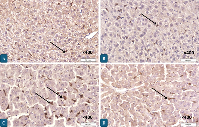
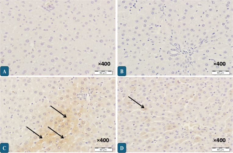
Tamoxifen (TAM) is extensively used to manage estrogen receptor-positive breast cancer. Despite its effectiveness, its administration can negatively impact various organs, including the liver. This research focused on the effects of TAM on the pyroptotic pathway in the liver and evaluated the potential of cardamom extract (CRDE) to lessen hepatic damage of TAM in female rats. Rats received 45 mg/kg of TAM injections for 10 days, while the groups treated with CRDE received 12 ml/kg of CRDE for 20 days, commencing 10 days before TAM administration. TAM exposure resulted in apparent degenerations in hepatic tissue with inflammatory cell infiltration and loss of architectures. Serum levels of liver enzymes including alanine aminotransferase, aspartate aminotransferase and alkaline phosphatase were elevated, along with hepatic oxidative stress, as shown by increased lipid peroxidation with lower levels of reduced glutathione. TAM caused inflammation in the liver tissue as indicated by higher levels of tumor necrosis factor-α and interleukin-6 as well as increased expression of CD68; a phagocytic Kupffer's cells marker. Additionally, the protein expression analysis revealed a high expression of pyroptotic markers including NLRP3-inflammasome, caspase-1, and gasdermin D. Conversely, CRDE treatment effectively neutralized the biochemical, histological, and protein expression alterations induced by TAM. In conclusion, CRDE demonstrated the potential to protect the liver from TAM-induced damage by regulating mechanisms involving oxidative damage, inflammation, and pyroptosis.
Keywords: CD68; Cardamom extract; Liver damage; Liver inflammation; Pyroptosis; Tamoxifen.
© 2025. The Author(s).
Conflict of interest statement
Declarations. Competing interests: The authors declare no competing interests. Animal ethics: All experimental procedures were reviewed and authorized by the Research Ethics Committee at King Saud University (Ethics Reference No: KSU-SE‐20‐75).
Figures


Similar articles
-
Protective role of thymoquinone against liver damage induced by tamoxifen in female rats.Can J Physiol Pharmacol. 2014 Aug;92(8):640-4. doi: 10.1139/cjpp-2014-0148. Epub 2014 May 26. Can J Physiol Pharmacol. 2014. PMID: 24941454
-
Zinc abrogates anticancer drug tamoxifen-induced hepatotoxicity by suppressing redox imbalance, NO/iNOS/NF-ĸB signaling, and caspase-3-dependent apoptosis in female rats.Toxicol Mech Methods. 2020 Feb;30(2):115-123. doi: 10.1080/15376516.2019.1669243. Epub 2019 Oct 1. Toxicol Mech Methods. 2020. PMID: 31532279
-
Pioglitazone attenuates tamoxifen-induced liver damage in rats via modulating Keap1/Nrf2/HO-1 and SIRT1/Notch1 signaling pathways: In-vivo investigations, and molecular docking analysis.Mol Biol Rep. 2023 Dec;50(12):10219-10233. doi: 10.1007/s11033-023-08847-x. Epub 2023 Nov 7. Mol Biol Rep. 2023. PMID: 37934372 Free PMC article.
-
Amelioration of tamoxifen-induced liver injury in rats by grape seed extract, black seed extract and curcumin.Indian J Exp Biol. 2010 Mar;48(3):280-8. Indian J Exp Biol. 2010. PMID: 21046982
-
Protective effects of cardamom aqueous extract against tamoxifen-induced pancreatic injury in female rats.Toxicol Res. 2023 Jun 27;39(4):721-737. doi: 10.1007/s43188-023-00198-w. eCollection 2023 Oct. Toxicol Res. 2023. PMID: 37779590 Free PMC article.
References
-
- Farrar, M. C. & Jacobs, T. F. In Tamoxifen (StatPearls Publishing, 2024). - PubMed
-
- El-Kashef, D. H. & El-Sheakh, A. R. Hepatoprotective effect of celecoxib against tamoxifen-induced liver injury via inhibiting ASK-1/JNK pathway in female rats. Life Sci.231, 116573 (2019). - PubMed
-
- Ribeiro, M. P. C., Santos, A. E. & Custódio, J. B. A. Mitochondria: the gateway for tamoxifen-induced liver injury. Toxicology323, 10–18 (2014). - PubMed
MeSH terms
Substances
Grants and funding
LinkOut - more resources
Full Text Sources
Medical
