. 2025 Feb 8;23(1):97.
doi: 10.1186/s12951-025-03191-7.

RBD-displaying OMV nanovaccine boosts immunity against SARS-CoV-2

Affiliations

RBD-displaying OMV nanovaccine boosts immunity against SARS-CoV-2

Rang Feng et al. J Nanobiotechnology. .

Abstract

Background: Since the emergence of SARS-CoV-2, the causative agent of COVID-19, the global health landscape has confronted an unprecedented and formidable challenge. The SARS-CoV-2 receptor-binding domain (RBD) is a key antigen in vaccine design. However, its low immunogenicity has been a hurdle, resulting in the production of minimal anti-RBD antibodies even when combined with alum adjuvant. Outer membrane vesicles (OMVs), secreted by Gram-negative bacteria, are nanospherical structures that can display or deliver antigens while also providing adjuvant activity through pathogen-associated molecular patterns (PAMPs).

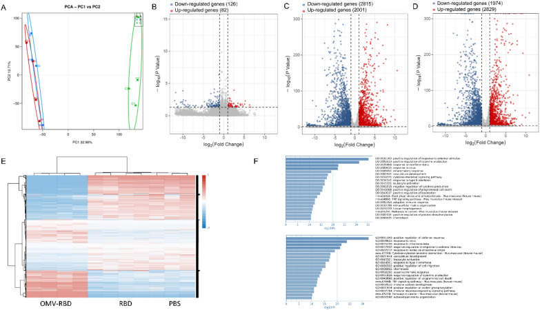
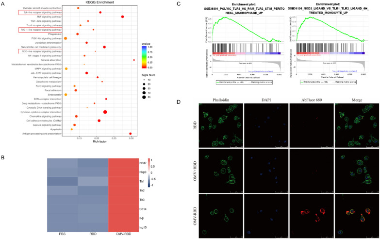
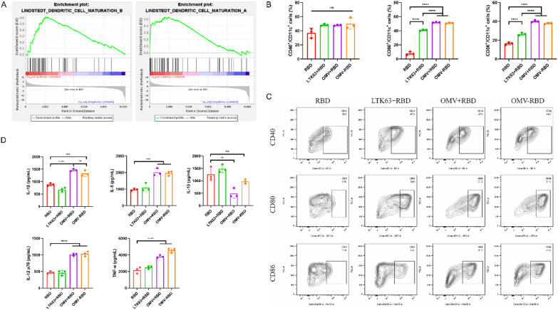
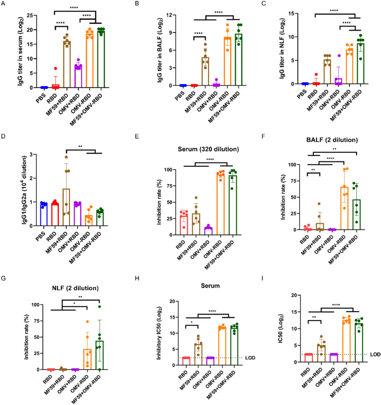
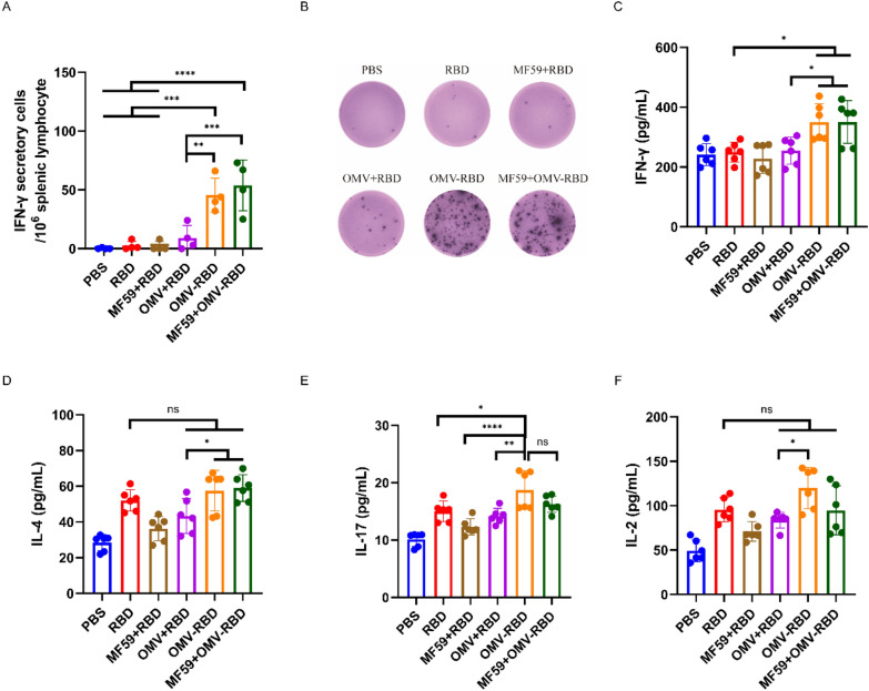
Results: In this study, we utilized the SpyTag (ST)/SpyCatcher (SC) bioconjugation system to couple OMV and SARS-CoV-2 RBD in vitro. We successfully prepared a 'plug-and-display' nanovaccine OMV-RBD, which demonstrated good safety profiles and promoted the uptake of antigens by DCs and the maturation of BMDCs by activating TLR3 and NOD2 signaling pathways. Both intranasal and intramuscular immunization with OMV-RBD vaccine elicited robust antigen-specific humoral and cellular immune responses. Importantly, the induced antibodies effectively inhibited the binding of RBD to human angiotensin-converting enzyme 2 (hACE2) and neutralized SARS-CoV-2 pseudoviruses.

Conclusions: This vaccine platform offers an alternative strategy for developing recombinant subunit vaccines against SARS-CoV-2, potentially enhancing immune responses and improving vaccine efficacy.


Conflict of interest statement

Declarations. Ethics approval and consent to participate: The Animal Ethics Committee of Army Medical University has approved all animal experiments. Trained experimentalists adhered to the 3R principle at all times during the experiment to avoid unnecessary suffering of the animals. Consent for publication: Not applicable. Competing interests: The authors declare no competing interests.

Figures

Fig. 1
Fig. 1
Preparation and characterization of OMV-RBD. A Schematic diagram of OMV-RBD preparation process. B Construction of pThio-HisA-ClyA-SC plasmid and pcDNA3.1-RBD-SpyTag plasmid. C The SDS-PAGE for screening the induction conditions. The ClyA bands at 55 kDa were different due to variations in IPTG induction concentrations and culture temperatures. D The reaction process and purification effect were monitored by Western blot. The predicted molecular weight of RBD-ST is 28 kDa. Lane 1: OMV-SC; Lane 2: RBD-ST; Lane 3: Reaction Product of OMV-SC and RBD-ST at a Mass Ratio of 30:1; Lane 4: Purified OMV-RBD after Ultracentrifugation. E Transmission electron microscopy of OMV-SC. F Transmission electron microscopy of OMV-SC
Fig. 2
Fig. 2
OMV-RBD activated the innate immune process in BMDCs. A Principal component analysis showed that PBS (red), RBD (blue) and OMV-RBD (green) treated samples in different groups. B Volcanic maps of DEGs in the RBD group and the PBS group (as control). Those marked in red are up-regulated genes (LFC > 1, P < 0.05), blue marked for down-regulated genes (LFC < −1, P < 0.05). The names of genes with significant and representative changes were highlighted. C Volcanic maps of differentially expressed genes (DEGs) in the OMV-RBD group and the PBS group (as control). D Volcanic maps of DEGs in in the OMV-RBD group and the RBD group (as control). E Heat maps of unsupervised cluster DEGs in samples. F Genes up-regulated in the OMV-RBD group compared to the PBS (top) or RBD (bottom) groups were clustered and annotated using Metscape, respectively, and visualized using bar graphs after collapsing redundant tags
Fig. 3
Fig. 3
OMV-RBD activated NOD2 and TLR3 on the surface of BMDCs. A Kyoto Encyclopedia of Genes and Genomes (KEGG) pathway analysis of up-regulated genes in OMV-RBD group vs. PBS group (as control). B Heatmap of the core genes in the selected pathway. Gene expression was measured by Transcripts Per Million (TPM) and represented by color. C Gene expression profile analysis by gene set enrichment analysis (GSEA) revealed significant enrichment of TLR3 and NOD2 signaling pathways related gene features of OMV-RBD up-regulated genes (P < 0.05). Y-axes indicate enrichment scores (top) and ranked list metric (bottom). X-axis bars represent individual genes of the indicated gene sets. D OMV-RBD can promote recognition and phagocytosis of dendritic cells. The confocal microscope images of BMDCs incubated with RBD, OMV + RBD and OMV-RBD respectively. The cytoskeleton was stained with TRITC phalloidin, and the nucleus was displayed by binding DAPI. RBD was labeled with Abfluor 680
Fig. 4
Fig. 4
OMV-RBD confered the ability of BMDCs to initiate adaptive immune responses. A Enrichment plots of Lindstedt DC maturation gene sets showing significant upregulation in OMV-RBD treated BMDCs. Representative plots C and statistical plots B showed the surface expression of CD40, CD80 and CD86 in CD11c + cell population after co-incubation of BMDCs with RBD, LTK63 + RBD, OMV + RBD and OMV-RBD respectively. D The cytokines in the supernatant of BMDCs culture were detected by ELISA. Data are expressed as mean ± S.D., n = 3. (*P < 0.05, **P < 0.01, ***P < 0.001, ****P < 0.0001)
Fig. 5
Fig. 5
Humoral response of intranasally immunized mice. A The levels of mouse sIgA antibody changed in vaginal wash post-immunization. B Specific sIgA titers in BALF. C Specific sIgA titers in NLF. D Specific IgG titers in BALF. E Specific IgG titers in NLF. F Specific sIgA titers in serum. G Specific IgG titers in serum. H The ratio of IgG1/IgG2a in serum. I hACE2 competitive inhibition rate of serum; (J) hACE2 competitive inhibition rate of NLF; K IC50 of hACE2 competitive inhibition in serum. L Serum pseudovirus neutralizing antibody levels. Data are expressed as mean ± S.D., n = 6. (*P < 0.05, **P < 0.01, ***P < 0.001, ****P < 0.0001)
Fig. 6
Fig. 6
Cellular response of intranasally immunized mice. A The number of RBD-specific IFN-γ secretory cells in 106 pulmonary lymphocytes. B Representative ELISpot results of pulmonary lymphocytes. C The number of RBD-specific IFN-γ secretory cells in 106 spleen lymphocytes. D Representative ELISpot results of spleen lymphocytes. E The concentration of IFN-γ. F The concentration of IL-2. G The concentration of IL-4. H The concentration of IL-17. Data are expressed as mean ± S.D., n = 6. (*P < 0.05, **P < 0.01, ***P < 0.001)
Fig. 7
Fig. 7
Humoral response of intramuscularly immunized mice. A Specific IgG titers in serum. B Specific IgG titers in BALF. C Specific IgG titers in NLF. D The ratio of IgG1/IgG2a in serum. E hACE2 competitive inhibition rate of serum; F hACE2 competitive inhibition rate of BALF; G hACE2 competitive inhibition rate of NLF. H IC50 of hACE2 competitive inhibition in serum. (I) Serum pseudovirus neutralizing antibody levels. Data are expressed as mean ± S.D., n = 6. (*P < 0.05, **P < 0.01, ****P < 0.0001)
Fig. 8
Fig. 8
Cellular response of intramuscularly immunized mice. A The number of RBD-specific IFN-γ secretory cells in 106 spleen lymphocytes. B Representative ELISpot results of spleen lymphocytes. C The concentration of IFN-γ. D The concentration of IL-2. E The concentration of IL-4. F The concentration of IL-17. Data are expressed as mean ± S.D., n = 6. (*P < 0.05, **P < 0.01, ***P < 0.001, ****P < 0.0001)

References

    1. Lundstrom K, Hromic-Jahjefendic A, Bilajac E, Aljabali AAA, Baralic K, Sabri NA, et al. COVID-19 signalome: Pathways for SARS-CoV-2 infection and impact on COVID-19 associated comorbidity. Cell Signal. 2023;101: 110495. - PMC - PubMed
    1. Bramble J, Fulk A, Saenz R, Agusto FB. Exploring the role of superspreading events in SARS-CoV-2 outbreaks. J Theor Biol. 2023;558: 111353. - PMC - PubMed
    1. Chen A, Wessler T, Gregory FM. Antibody protection from SARS-CoV-2 respiratory tract exposure and infection. J Theor Biol. 2023;557: 111334. - PMC - PubMed
    1. Castells MC, Phillips EJ. Maintaining safety with SARS-CoV-2 Vaccines. N Engl J Med. 2021;384(7):643–9. - PMC - PubMed
    1. Le Thanh T, Andreadakis Z, Kumar A, Gomez Roman R, Tollefsen S, Saville M, et al. The COVID-19 vaccine development landscape. Nat Rev Drug Discov. 2020;19(5):305–6. - PubMed

MeSH terms

LinkOut - more resources