

Variations in intracranial arterial anatomy of the circle of Willis and their association with arteriosclerosis in patients with ischemic cerebrovascular disease
- PMID: 39924650
- PMCID: PMC12264298
- DOI: 10.1177/17474930251322678
Variations in intracranial arterial anatomy of the circle of Willis and their association with arteriosclerosis in patients with ischemic cerebrovascular disease
Abstract
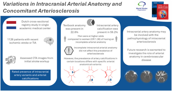
Introduction: An estimated 20-31% of all people are born with a textbook anatomical configuration of the intracranial arteries comprising the Circle of Willis. Individuals with specific anatomical variants may be at elevated risk of intracranial arteriosclerosis, and possibly its sequelae of stroke and dementia, as the distribution of blood flow and pressure is known to be different in variants with missing arteries or arterial segments. Therefore, we studied the association of anatomical variation of intracranial arteries with arteriosclerosis.
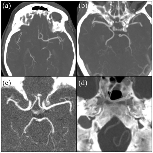
Methods: Between December 2005 and October 2010, 1126 patients (mean age: 62.3 (SD: ±14.0) years, 48.0% female) were recruited, 59.9% of whom had ischemic stroke and 40.1% a transient ischemic attack (TIA). Within the routine diagnostic work-up for stroke, patients underwent cranial computed tomography (CT) angiography. These images enabled a detailed visualization of intracranial arteries, which allowed for the assessment of the anatomical configuration of the cerebral arteries, the anterior and posterior communicating arteries, the internal carotids, and the vertebrobasilar arteries. In addition, these images facilitated the identification of intracranial arterial calcifications, the defining feature of intracranial arteriosclerosis. Binomial logistic regression models adjusting for age, sex, and ethnicity were constructed to assess associations between intracranial artery variations and presence of intracranial arterial calcifications.
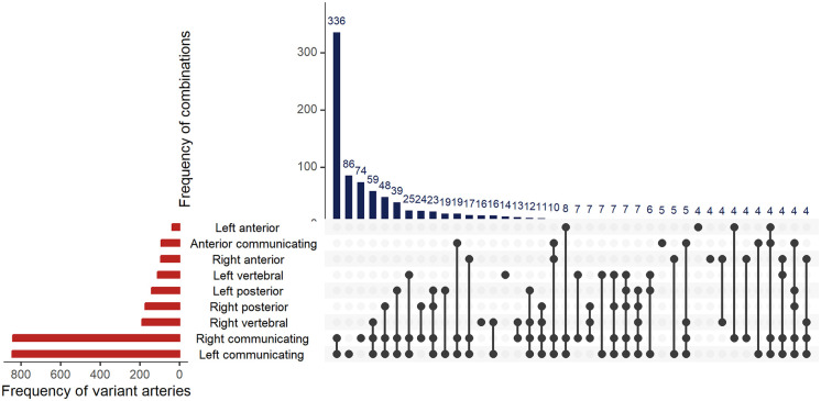
Results: An incomplete Circle of Willis, defined by aplasia of any arterial segment, was present in 875 (77.7%) patients. The most common variation found was aplasia of the right posterior communicating artery, in 52.0% of patients. Men more often presented with an incomplete anatomy as compared to women (adjusted odds ratio: 1.36 (95% CI = 1.02-1.81)). Intracranial artery calcification was present in 59.2% of patients. Incompleteness of the intracranial arteries was not associated with the presence of any intracranial artery calcification (0.95 (0.68-1.34)). However, specific variants were associated with specific locations of intracranial artery calcification: The prevalence of vertebrobasilar artery calcification was lower among those with fetal-type posterior cerebral artery compared to individuals with a normal posterior cerebral artery (0.61 (0.38-0.99)). The prevalence of vertebrobasilar artery calcification was higher among those with a-/hypoplasia of both posterior communicating arteries as compared to those with normal posterior communicating arteries (1.63 (1.00-2.66)). Furthermore, patients with a-/hypoplastic left A1-segments had a higher prevalence of right internal carotid artery calcification as compared to people with a normal left A1-segment (2.30 (1.00-5.26)).
Conclusion: The prevalence of arteriosclerosis in the intracranial arteries on CT imaging varies among patients with certain anatomical variants of the intracranial arterial system. Specifically, arteriosclerosis in the right internal carotid artery and the vertebrobasilar arteries was more frequently observed in patients who had an a-/hypoplastic left anterior cerebral artery or a-/hypoplasia of both posterior communicating arteries, respectively. In addition, arteriosclerosis was less frequently observed among vertebrobasilar arteries of patients with a fetal-type posterior cerebral artery. Future longitudinal research is warranted regarding the anatomical configuration of intracranial arteries and the development of intracranial arteriosclerosis, as this line of research may reveal a novel group of people at elevated risk of cerebrovascular disease.
Keywords: Stroke; anatomy; arteriosclerosis; intracranial arterial diseases; vascular calcification.
Conflict of interest statement
Declaration of conflicting interestsThe author(s) declared no potential conflicts of interest with respect to the research, authorship, and/or publication of this article.
Figures
Similar articles
-
Arterial brain calcium (ABC) volume - A novel radiological marker of atherosclerotic risk and future stroke risk on non-contrast CT.J Stroke Cerebrovasc Dis. 2025 Aug;34(8):108359. doi: 10.1016/j.jstrokecerebrovasdis.2025.108359. Epub 2025 May 26. J Stroke Cerebrovasc Dis. 2025. PMID: 40436256
-
Sex-Specific Association between Anatomic Variants of the Circle of Willis and Intracranial Aneurysms in Individuals with a Familial Predisposition.AJNR Am J Neuroradiol. 2025 Aug 1;46(8):1542-1547. doi: 10.3174/ajnr.A8690. AJNR Am J Neuroradiol. 2025. PMID: 39909568
-
Automated devices for identifying peripheral arterial disease in people with leg ulceration: an evidence synthesis and cost-effectiveness analysis.Health Technol Assess. 2024 Aug;28(37):1-158. doi: 10.3310/TWCG3912. Health Technol Assess. 2024. PMID: 39186036 Free PMC article.
-
Modelling blood flow in the circle of Willis in continuous flow left ventricular assist devices: possible relevance to strokes.Indian J Thorac Cardiovasc Surg. 2025 Feb;41(2):148-155. doi: 10.1007/s12055-024-01806-6. Epub 2024 Sep 12. Indian J Thorac Cardiovasc Surg. 2025. PMID: 39822874
-
Signs and symptoms to determine if a patient presenting in primary care or hospital outpatient settings has COVID-19.Cochrane Database Syst Rev. 2022 May 20;5(5):CD013665. doi: 10.1002/14651858.CD013665.pub3. Cochrane Database Syst Rev. 2022. PMID: 35593186 Free PMC article.
References
-
- Jones JD, Castanho P, Bazira P, Sanders K. Anatomical variations of the circle of Willis and their prevalence, with a focus on the posterior communicating artery: a literature review and meta-analysis. Clin Anat 2021; 34: 978–990. - PubMed
-
- Hendrikse J, van Raamt AF, van der Graaf Y, Mali WP, van der Grond J. Distribution of cerebral blood flow in the circle of Willis. Radiology 2005; 235: 184–189. - PubMed
-
- Liebeskind DS, Caplan LR. Intracranial arteries—anatomy and collaterals. Front Neurol Neurosci 2016; 40: 1–20. - PubMed
MeSH terms
LinkOut - more resources
Full Text Sources
Medical
