Review
. 2025 Feb 3:18:1479-1495.
doi: 10.2147/JIR.S504184. eCollection 2025.

The Mechanisms of Sepsis Induced Coagulation Dysfunction and Its Treatment

Affiliations
Review

The Mechanisms of Sepsis Induced Coagulation Dysfunction and Its Treatment

Lei Zhu et al. J Inflamm Res. .

Abstract

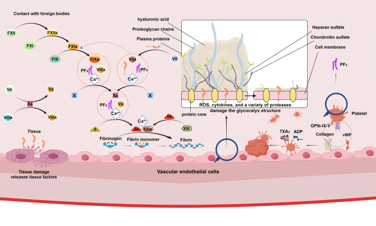
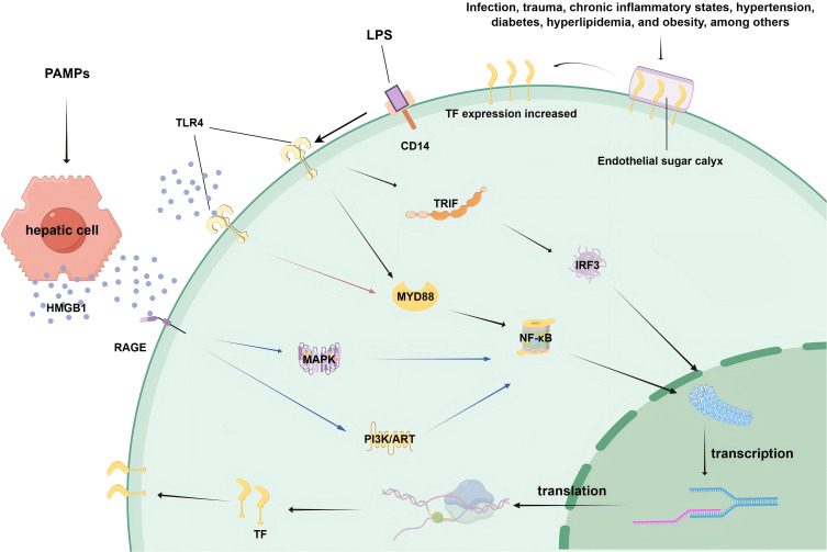
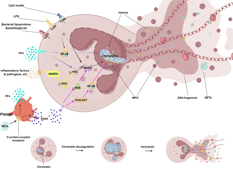
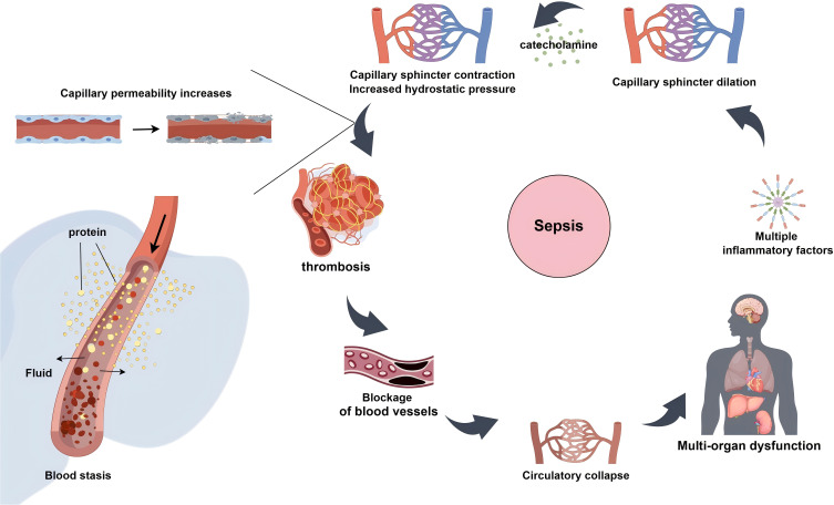
Sepsis is a critical condition characterized by organ dysfunction due to a dysregulated response to infection that poses significant global health challenges. Coagulation dysfunction is nearly ubiquitous among sepsis patients. Its mechanisms involve platelet activation, coagulation cascade activation, inflammatory reaction imbalances, immune dysregulation, mitochondrial damage, neuroendocrine network disruptions, and endoplasmic reticulum (ER) stress. These factors not only interact but also exacerbate one another, leading to severe organ dysfunction. This review illustrates the mechanisms of sepsis-induced coagulopathy, with a focus on tissue factor activation, endothelial glycocalyx damage, and the release of neutrophil extracellular traps (NETs), all of which are potential targets for therapeutic interventions.

Keywords: coagulation dysfunction; sepsis; thrombosis.


Conflict of interest statement

The authors report no conflicts of interest in this work.

Figures

Figure 1
Figure 1
Activation of Platelets, Coagulation Cascade, and Endothelial Glycocalyx Damage. The figure on the left shows the activation of the coagulation cascade, with multiple coagulation factors contributing to the synthesis of fibrin; the middle figure depicts damage to the endothelial glycocalyx, which becomes thinner and sparser, leading to increased exposure of TF; and the right figure side illustrates the activation and aggregation of platelets.
Figure 2
Figure 2
Expression of TF. The process by which endothelial glycocalyx damage leads to increased exposure of TF. In response to stimuli such as infection, trauma, and chronic inflammation, monocytes/macrophages and endothelial cells activate signaling pathways via HMGB1 and PAMP receptors (eg, TLR4 and RAGE), resulting in increased expression of transcription factors.
Figure 3
Figure 3
The formation of the NETs. Signaling pathways and cellular processes involved in the formation and release of NETs during the immune response. This highlights the role of TLRs in recognizing bacterial components, the NF‒κB pathway triggered by TLR signaling, and the PKC/MAPK pathway involved in chromatin remodeling. The NADPH oxidase complex contributes to ROS formation, whereas the PI3K/AKT pathway contributes to chromatin decondensation. MPO and histone DNA fragments are also involved in this process, leading to the formation of NETs. PF4 and PDGF interact with neutrophils, and inflammatory factors and pathogens act as stimuli for NETs.
Figure 4
Figure 4
Progression of thrombosis in the context of sepsis. Sepsis induces the release of multiple inflammatory mediators, which damage vascular endothelial cells, ultimately causing microvascular constriction, increased capillary permeability, and elevated hydrostatic pressure. This cascade leads to the leakage of fluid and proteins, blood stasis within the microcirculation, and consequently, the formation of thrombi. This sequence can culminate in microcirculatory dysfunction and multiorgan failure.

Similar articles

Cited by

References

    1. Fleischmann-Struzek C, Mellhammar L, Rose N, et al. Incidence and mortality of hospital- and ICU-treated sepsis: results from an updated and expanded systematic review and meta-analysis. Intensive Care Med. 2020;46(8):1552–1562. doi:10.1007/s00134-020-06151-x - DOI - PMC - PubMed
    1. Rudd KE, Johnson SC, Agesa KM, et al. Global, regional, and national sepsis incidence and mortality, 1990-2017: analysis for the global burden of disease study. Lancet. 2020;395(10219):200–211. doi:10.1016/S0140-6736(19)32989-7 - DOI - PMC - PubMed
    1. Xie J, Wang H, Kang Y, et al. The epidemiology of sepsis in Chinese ICUs: a national cross-sectional survey. Crit Care Med. 2020;48(3):e209–e218. doi:10.1097/CCM.0000000000004155 - DOI - PubMed
    1. Singer M, Deutschman CS, Seymour CW, et al. The third international consensus definitions for sepsis and septic shock (Sepsis-3). JAMA. 2016;315(8):801–810. doi:10.1001/jama.2016.0287 - DOI - PMC - PubMed
    1. Schmoch T, Möhnle P, Weigand MA, et al. The prevalence of sepsis-induced coagulopathy in patients with sepsis - a secondary analysis of two German multicenter randomized controlled trials. Ann Intens Care. 2023;13(1):3. doi:10.1186/s13613-022-01093-7 - DOI - PMC - PubMed

LinkOut - more resources