. 2025 Jan 22:10:100271.
doi: 10.1016/j.jtauto.2025.100271. eCollection 2025 Jun.

Tofacitinib downregulates JAK1 and JAK3 on human intestinal monocytes and macrophages without affecting dendritic cells phenotype or function

Affiliations

Tofacitinib downregulates JAK1 and JAK3 on human intestinal monocytes and macrophages without affecting dendritic cells phenotype or function

Elisa Arribas-Rodríguez et al. J Transl Autoimmun. .

Abstract

Background: Ulcerative colitis (UC) is an inflammatory disorder of the gastrointestinal tract. Although Tofacitinib, which inhibits the JAK1 and JAK3 signalling pathway, is approved to treat patients with UC, its specific mechanism of action remain elusive. Given the central role that conventional dendritic cells (cDC) elicit in gut homeostasis, we hypothesised that Tofacitinib acts modulating cDC function in UC.

Methods: Human biopsies were obtained from colon of controls, and patients with UC (active and quiescent). Lamina propria mononuclear cells (LPMC) were ex-vivo cultured in the presence/absence of Tofacitinib. The specific effect elicited over human intestinal cDC, monocytes and macrophages was assessed by flow cytometry. cDC were also enriched following Tofacitinib conditioning in order to assess its effect over naïve T-cells.

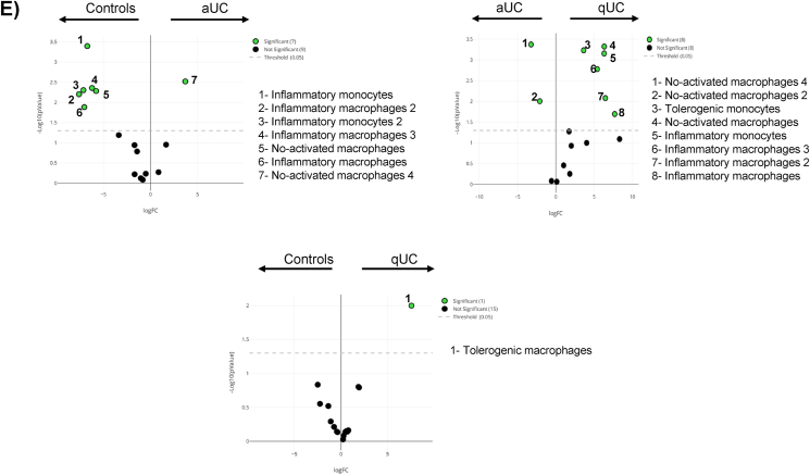
Results: Several human intestinal cDC, monocyte and macrophage subsets can be found in the human colon, with these cells being more similar between controls and patients with qUC referred to patients with aUC. Following ex-vivo culture, Tofacitinib downregulated JAK1 expression on intestinal monocytes from patients with both active and quiescent UC. As for macrophages, JAK1 was decreased on patients with active UC while JAK was downregulated on macrophages from patients with quiescent disease. Tofacitinib did not modulate the phenotype or function of human intestinal cDC.

Conclussion: Tofacitinib does not modulate the phenotype and function of human intestinal cDC in UC. On the contrary, it displays a differential capacity to modulate intestinal monocyte and macrophage phenotype. Future studies should address whether it also translates into a differential function of these cells.

Keywords: Dendritic cells; Intestine; Macrophages; Tofacitinib; Ulcerative colitis.


Conflict of interest statement

The authors declare that they have no known competing financial interests or personal relationships that could have appeared to influence the work reported in this paper.

Figures

Fig. 1
Fig. 1
Unsupervised analysis of human intestinal antigen presenting cells. A) Total myeloid antigen presenting cells (mAPC) were identified within singlet viable leukocytes as in Supplementary Fig. 3, and analyzed with a Uniform Manifold Approximation and Projection (UMAP) on resting conditions (n = 30). B) Expression intensities of the analyzed markers represented with a color code based on the intensity where red represent higher expression and blue, lower expression. C) Heatmap displaying the intensity levels of each identified cluster within the three cohorts. D) All 16 clusters were overlaid on the UMAP projection using a specific color and number as shown in the legend. E) Volcano plots comparing the different clusters among the 3 study groups highlighting in green those with statistically significant differences.
Fig. 1
Fig. 1
Unsupervised analysis of human intestinal antigen presenting cells. A) Total myeloid antigen presenting cells (mAPC) were identified within singlet viable leukocytes as in Supplementary Fig. 3, and analyzed with a Uniform Manifold Approximation and Projection (UMAP) on resting conditions (n = 30). B) Expression intensities of the analyzed markers represented with a color code based on the intensity where red represent higher expression and blue, lower expression. C) Heatmap displaying the intensity levels of each identified cluster within the three cohorts. D) All 16 clusters were overlaid on the UMAP projection using a specific color and number as shown in the legend. E) Volcano plots comparing the different clusters among the 3 study groups highlighting in green those with statistically significant differences.
Fig. 2
Fig. 2
Tofacitinib effect over human intestinal antigen presenting cells. Total lamina propria mononuclear cells (LPMC) from controls, were ex-vivo cultured in resting conditions (Basal), as well as with 100 ng/ml of LPS in the presence/absence of 100 nM Tofacitinib. Total monocytes, macrophages and conventional dendritic cells (cDC) were identified as in Supplementary Fig. 1, as assessed for the expression of CXCR3, IL-10, IL-15, IL-1β, IL-6, TLR2, TLR4, TNFα, JAK1 and JAK3 based on their respective fluorescence minus one (FMO) controls as in Supplementary Fig. 2. Two-way ANOVA was applied P-values <0.05 were considered significant (∗p < 0.05; ∗∗p < 0.01; ∗∗∗p < 0.001; ∗∗∗∗p < 0.0001).
Fig. 3
Fig. 3
Tofacitinib modulation of human intestinal antigen presenting cells from patients with active ulcerative colitis. Total lamina propria mononuclear cells (LPMC) from patients with active ulcerative colitis were ex-vivo cultured in resting conditions (Basal) as well as with 100 nM Tofacitinib. Subsequent expression of CXCR3, IL-10, IL-15, IL-1β, IL-6, TLR2, TLR4, TNFα, JAK1 and JAK3 on monocytes, macrophages and conventional dendritic cells (cDC) was determined as in Fig. 2. Two-way ANOVA was applied panel P-values <0.05 were considered significant (∗p < 0.05; ∗∗p < 0.01; ∗∗∗p < 0.001; ∗∗∗∗p < 0.0001).
Fig. 4
Fig. 4
Tofacitinib effect over human intestinal antigen presenting cells from patients with quiescent ulcerative colitis. Total lamina propria mononuclear cells (LPMC) from patients with quiescent ulcerative colitis were ex-vivo cultured in resting conditions (Basal) as well as with 100 nM Tofacitinib. Subsequent expression of CXCR3, IL-10, IL-15, IL-1β, IL-6, TLR2, TLR4, TNFα, JAK1 and JAK3 on monocytes, macrophages and conventional dendritic cells (cDC) was determined as in Fig. 2. Two-way ANOVA was applied P-values <0.05 were considered significant (∗p < 0.05; ∗∗p < 0.01; ∗∗∗p < 0.001; ∗∗∗∗p < 0.0001).
Fig. 5
Fig. 5
Tofacitinib effect over the immunostimulatory capacity of human intestinal conventional dendritic cells. A) Total lamina propria mononuclear cells were ex-vivo cultured in resting conditions (Basal), as well as with 100 ng/ml of LPS in the presence/absence of 100 nM Tofacitinib. Total conventional dendritic cells (cDC) were subsequently sorted and co-cultured with allogeneic cell-trace violet labelled naïve T-cells. cDC stimulatory capacity over total T-cells was determined, as well as B) the stimulatory capacity specifically elicited over both CD4 and CD8. The acquired phenotype (Treg: FOXP3+IL-10+; Th1: Tbet+IFNᵞ+; Th17: RORᵞt+IL17+ is displayed in C). Two-way ANOVA was applied in all cases. P-values <0.05 were considered significant (∗p < 0.05; ∗∗p < 0.01; ∗∗∗p < 0.001; ∗∗∗∗p < 0.0001).

References

    1. Mowat A.M.I. Anatomical basis of tolerance and immunity to intestinal antigens. Nat. Rev. Immunol. 2003;3:331–341. doi: 10.1038/nri1057. - DOI - PubMed
    1. Zhang Y.Z., Li Y.Y. Inflammatory bowel disease: pathogenesis. World J. Gastroenterol. 2014;20:91–99. doi: 10.3748/wjg.v20.i1.91. - DOI - PMC - PubMed
    1. Atreya R., Neurath M.F. IBD pathogenesis in 2014 : molecular pathways controlling barrier function in IBD. Nat. Rev. Gastroenterol. Hepatol. 2015;12:37–38. doi: 10.1038/nrgastro.2014.201. - DOI - PubMed
    1. Boland B.S., Sandborn W.J., Chang J.T. Update on Janus kinase antagonists in inflammatory bowel disease. Gastroenterol Clin North Am. 2014;43:603–617. doi: 10.1016/j.gtc.2014.05.011. - DOI - PMC - PubMed
    1. Xin P., Xu X., Deng C., Liu S., Wang Y., Zhou X., et al. The role of JAK/STAT signaling pathway and its inhibitors in diseases. Int Immunopharmacol. 2020;80 doi: 10.1016/j.intimp.2020.106210. - DOI - PubMed

LinkOut - more resources