. 2025 Feb 4;9(1):010901.
doi: 10.1063/5.0220516. eCollection 2025 Mar.

Biosensors integrated within wearable devices for monitoring chronic wound status

Affiliations

Biosensors integrated within wearable devices for monitoring chronic wound status

Sabine Szunerits et al. APL Bioeng. .

Abstract

Slowly healing wounds significantly affect the life quality of patients in different ways, due to constant pain, unpleasant odor, reduced mobility up to social isolation, and personal frustration. While remote wound management has become more widely accepted since the COVID-19 pandemic, delayed treatment remains frequent and results in several wound healing related complications. As inappropriate management of notably diabetic foot ulcers is linked to a high risk of amputation, effective management of wounds in a patient-centered manner remains important to be implemented. The integration of diagnostic devices into wound bandages is under way, owing to advancements in materials science and nanofabrication strategies as well as innovation in communication technologies together with machine learning and data-driven assessment tools. Leveraging advanced analytical approaches around local pH, temperature, pressure, and wound biomarker sensing is expected to facilitate adequate wound treatment. The state-of-the-art of time-resolved monitoring of the wound status by quantifying key physiological parameters as well as wound biomarkers' concentration is presented herewith. A special focus will be given to smart bandages with on-demand delivery capabilities for improved wound management.


Conflict of interest statement

The authors have no conflicts to disclose.

Figures

FIG. 1.
FIG. 1.
Wound care decisions can be made using the TIMERS concept. (A) pH, temperature, moisture, and biomarkers for continuous monitoring. (B) Elements of the TIMERS concept [created with BioRender software (BioRender.com)]. (C) Details of the TIMERS concept: boxes specify the state of the wound assessment and arrows indicate the action/treatment to be followed. Epithelium tissue appears as pink in the final stage of healing, red when healthy tissue is formed in the remodeling phase, brown due to devitalized tissue made of dead cells (slough), while necrotic tissue is black. Inflammation can persist due to infection requiring wound cleaning and treatment depending on the wound state. Moisture is essential to healing, and the treatment focuses on retaining and containing it within the wound bed. The measurement of the size and depth of the wound together with the identification of the edges is a key step in clinical assessment. Healing outcome and tissue repair depend on the chosen treatment approaches, based on expert experience. This Figure was constructed based on Refs. and .
FIG. 2.
FIG. 2.
Advances in pH sensors for wounds. (A) (i) Synthesis of O-CDs. (ii) Transmission electron microscopy image and corresponding electron diffraction pattern of O-CDs. (iii) Conceptual view of the practical application. (iv) Change in color under white light irradiation as well as fluorescence images under natural, 365 and 254 nm excitations of O-CDs/MCC at various pH values. [Reprint with permission from Yang et al., Small 15, 1902823 (2019). Copyright 2019 John Wiley and Sons]. (B) (i) Schematics of the pH sensor array. (ii) Cross section of embedded wound sensor. (iii) Sensor response to pH changes. [Reprint with permission from Rahimi et al., Sens. Actuators, B 229, 609–617 (2016). Copyright 2016 Elsevier]. (C) (i) Schematics of textile PEDOT:PSS/IrOx pH sensor. (ii) Wound bandage sensor design. (iii) Sensor time response in universal buffer solution; inset is the calibration plot. (iv) Response of the sensor in simulated wound exudate; inset is the calibration plot. (v) Signal recovery in simulated wound exudate. [Reprint with permission from Mariani et al., ACS Sens. 6, 2366–2377 (2021). Copyright 2021 Authors, licensed under a Creative Commons Attribution (CC BY) license].
FIG. 3.
FIG. 3.
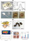
Temperature sensing related to wounds. (A) (i) Design of the temperature sensors based on a spiral coil inductor (L) and PEG-based capacitor (C). (ii) Shift of the LC-resonance peak vs. temperature. (iii) In vivo subcutaneous temperature measurements were taken with sensors implanted in rats. (iv) Details of the implantation procedure. [Reprint with permission from Lu et al., Adv. Healthcare Mater. 9, 2000942 (2020). Copyright 2020 John Wiley and Sons]. (B) (i) Flexible circuit board and conducting adhesive hydrogel integrated onto the bandage. (ii) Photographs of mice wearing the wound bandage. (iii) Infrared image of a mouse wearing the smart bandage (top) and traces of wirelessly sensed temperature and impedance (bottom). (iv) Photographs and quantitative comparison of wounds infected with E. coli, with and without stimulation. [Reprint with permission from Jiang et al., Nat. Biotechnol. 41, 652–662 (2023). Copyright 2023 Springer Nature].
FIG. 4.
FIG. 4.
Detection of wound biomarkers. (A) Top: The various components of the liquid bandage formulation: New-Skin® ethanol-nitrocellulose matrix, oxygen-sensing metalloporphyrin, and fluorescein dye. Bottom: Protocol for bandage fabrication. [Reprint with permission from Marks et al., Sci. Adv. 6, eabd1061 (2020). Copyright 2020 American Association for the Advancement of Science]. (B) Picture of Absorbest Fuktsensor on wound dressing. Image of the dressing with conductor coil and display placed outside. [Reprint with permission from Henricson et al., Skin Res. Technol. 27, 918–924 (2021). Copyright 2021 Authors, licensed under a Creative Commons Attribution (CC BY) license]. (C) OECT working mechanism for potentiostatic UA quantification. [Reprint with permission from Arcangeli et al., ACS Sens. 8, 1593–1608 (2023). Copyright 2023, American Chemical Society]. (D) Multianalyte skin sensor. [Reprint with permission from Gao et al., Sci. Adv. 7, eabg9614 (2021). Copyright 2021 Authors, licensed under a Creative Commons Attribution (CC BY) license].
FIG. 5.
FIG. 5.
Stretchable skin electronics. Flexible and transparent electronics allow simultaneous monitoring of biomarkers and visual inspection. (i) Descriptive diagram of a transistor array manufactured from intrinsically stretchable and transparent materials. (ii) An array of 108 stretchable transistors attached conformably to a fingertip. (iii) An array of 6300 stretchable transistors attached conformably to an inner wrist. (iv) An array of stretchable transistors attached conformably to a bent wrist. [Reproduced with permission from Wang et al., Nature 555, 83–88 (2018). Copyright 2018 Nature].
FIG. 6.
FIG. 6.
Artificial intelligence in wound management. (A) Artificial intelligence architectures in image-based wound management. (i) Wound segmentation based on Dual-Phase Hyperactive UNet. (ii) Wound detection based on YOLOv8. [Reprint with permission of Shah et al., Healthcare 11, 2840 (2023). Copyright 2023 Authors, licensed under a Creative Commons Attribution (CC BY) license]. (B) Multimodal artificial intelligence architecture for the management of chronic wounds employing disparate health databases (i), different types of data fusion models (ii), and decision/prediction multimodal algorithms (iii). [Reprint with permission of Kline et al., npj Digital Med. 5, 171 (2022). Copyright 2022 Authors, licensed under a Creative Commons Attribution (CC BY) license].

Similar articles

References

    1. London N. J. M. and Donnelly R., “ Ulcerated lower limb,” BMJ 320, 1589 (2000).10.1136/bmj.320.7249.1589 - DOI - PMC - PubMed
    1. Ellis S., Patel M., Koshchak E., and Lantis II J., “ Location of lower-extremity diabetic foot ulcers with concomitant arterial or venous disease,” Wounds International 11, 20–23 (2020).
    1. Alves P. J., Barreto R. T., Barrois B. M., Gryson L. G., Meaume S., and Monstrey S. J., “ Update on the role of antiseptics in the management of chronic wounds with critical colonisation and/or biofilm,” Int. Wound J. 18, 342–358 (2021).10.1111/iwj.13537 - DOI - PMC - PubMed
    1. Becker S. L., Kody S., Fett N. M., Hines A., Alavi A., and Ortega-Loayza A. G., “ Approach to the atypical wound,” Am. J. Clin. Dermatol. 25, 559–584 (2024).10.1007/s40257-024-00865-0 - DOI - PubMed
    1. Lumbers M., “ TIMERS: Undertaking wound assessment in the community,” Br. J. Community Nurs. 24, S22 (2019).10.12968/bjcn.2019.24.Sup12.S22 - DOI - PubMed