. 2025 Feb 4:18:161-177.
doi: 10.2147/OTT.S487126. eCollection 2025.

Crosstalk of SPINK4 Expression With Patient Mortality, Immunotherapy and Metastasis in Pan-Cancer Based on Integrated Multi-Omics Analyses

Affiliations

Crosstalk of SPINK4 Expression With Patient Mortality, Immunotherapy and Metastasis in Pan-Cancer Based on Integrated Multi-Omics Analyses

Xiuhua Cao et al. Onco Targets Ther. .

Abstract

Background: Cancer remains a major global health challenge, with early detection and prompt treatment being crucial for reducing mortality rates. The SPINK4 has been linked to the development of several tumors, and there is growing evidence of its involvement. However, its specific functions and effects in different cancer types remain unclear.

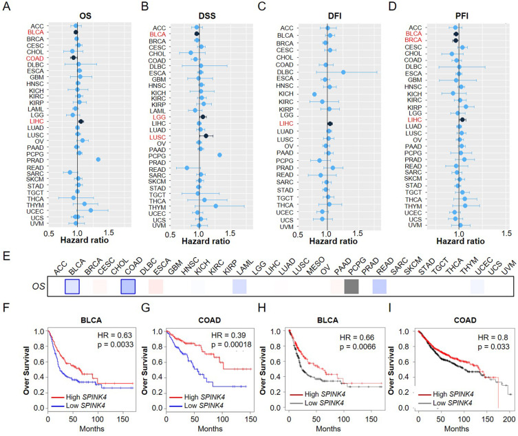
Methods: The association between SPINK4 expression levels and tumor progression was investigated and confirmed using the TCGA dataset. Kaplan-Meier curves were utilized to examine the correlation between SPINK4 expression with survival outcomes in pan-cancer patients. The Pearson method was employed to investigate the association of SPINK4 expression with the tumor microenvironment, stemness score, immunoinfiltrating subtype, and chemotherapy sensitivity in human different cancer types. Wound healing and Transwell assays were performed to confirm the roles of the model gene in colon adenocarcinoma cells.

Results: The expression of SPINK4 shows heterogeneity across pan-cancer tissues, and is closely associated with poor prognosis, immune cell invasion, tumor cell resistance, and tumor metastasis in a various human cancer. Mutation of SPINK4 hold significant predictive value for poor prognosis of pan-cancer patients. In addition, SPINK4 expression was significantly correlated with the tumor microenvironment (stromal cells and immune cells) and stemness score (DNAss and RNAss) in human pan-cancer tissues, particularly in BLCA and COAD. Single-cell sequencing analysis showed that SPINK4 is mainly expressed in endothelial cells in BLCA and in malignant cells in COAD. Drug resistance analysis showed a significant association between SPINK4 expression and sensitivity to several cancer chemotherapy drugs. Importantly, overexpression of SPINK4 promoted the metastasis of colon cancer cell lines (HCT116 and RKO), whereas SPINK4 knockout markedly inhibited their metastasis.

Conclusion: These findings reveal the crucial role of SPINK4 in the pan-cancer process and may have significant implications for the diagnosis and treatment of cancer in the future.

Keywords: drug sensitivity; genetic alteration; immune infiltration; pan-cancer analysis; single-cell sequencing.


Conflict of interest statement

The authors declare that they have no competing interests in this work.

Figures

Figure 1
Figure 1
Expression level of SPINK4 in pan-cancer. (A) Summary of mRNA and protein expression. (B) The protein structure and subcellular localization of SPINK4 (C) SPINK4 expression status in different types of cancers from TCGA (D) The expression level of SPINK4 in tumor and normal tissues via TIMER 2.0. (E) Differences in SPINK4 expression between normal, tumor, and metastatic tissues among COAD, ESCA, and KIRC. (F) The expression of SPINK4 in different types of cancers (BLCA, COAD, ESCA, KIRP, LIHC, THCA, and UCEC) were analyzed according to pathological stage (stage I, stage II, stage III, and stage IV). The p < 0.05 was considered to be statistically significant. *p < 0.05, **p < 0.01, ***p < 0.001.
Figure 2
Figure 2
Correlation between SPINK4 expression and overall survival in patients with different cancer types. (A) Cox analysis of the correlation between SPINK4 expression and OS in different cancer types. (B) The correlation of SPINK4 expression with DSS in different cancer types. (C) The correlation between SPINK4 expression and DFI in different cancer types. (D) Cox analysis of the correlation of SPINK4 expression with PFI in different cancer types. (E) Using GEPIA2 to construct an overall survival (OS) map of SPINK4 expression. (F) Kaplan–Meier survival curves (OS) of patients with high and low LDHA expression in BLCA. (G) Kaplan–Meier survival curves (OS) of patients with high and low LDHA expression in COAD. (H) Overall survival curves comparison of high or low expression of SPINK4 in BLCA by using Kaplan-Meier Plotter database. (I) Overall survival curves comparison of high or low expression of SPINK4 in COAD by using Kaplan-Meier Plotter database.
Figure 3
Figure 3
Analysis of SPINK4 genetic alterations in different types of cancers. (A) The frequency of SPINK4 mutations with mutation type across TCGA cancers by cBioPortal. (B) Oncoprint of SPINK4 alterations in cancer cohorts. (C-F) The associations of pan-cancer SPINK4 mutation status with OS, DSS, DFS and PFS by cBioPortal.
Figure 4
Figure 4
Correlation of SPINK4 expression with tumor microenvironment and stemness score in different types of cancers. (A) The correlation of SPINK4 expression with stromalscore, immunscore and ESTIMATEscore. (B) The correlation of SPINK4 expression with DNAss and RNAss. (C) The expression of SPINK4 correlated with stromal score, immune scores, and ESTIMATE scores in BLCA. (D) The expression of SPINK4 correlated with DNAss and RNAss in BLCA. (E) The expression of SPINK4 correlated with stromal score, immune scores, and ESTIMATE scores in COAD. (F) The expression of SPINK4 correlated with DNAss and RNAss in COAD. Gray brown background indicates no correlation between the gene expression and the corresponding index (p >0.05). Light background indicates that the gene is significantly correlated with the corresponding index (p <0.05). R represents correlation value, R >0 means positive correlation, R <0 means negative correlation.
Figure 5
Figure 5
Association of SPINK4 expression with immune infiltration subtypes in BLCA and COAD. (A). One-way analysis of variance was performed to test the correlation between SPINK4 expression and BLCA immune infiltration subtypes. (B) One-way analysis of variance was performed to test the correlation between SPINK4 expression and COAD immune infiltration subtypes. (C) Diagnostic value of SPINK4 expression in BLCA. (D) Diagnostic value of SPINK4 expression in COAD. (E) Association of SPINK4 expression with different immune cell in BLCA, including B cells, CD4 T-cells, CD8 T-cells, neutrophil, macrophage and dendritic cells. (F) Association of SPINK4 expression with different immune cell in COAD, including B cells, CD4 T-cells, CD8 T-cells, neutrophil, macrophage and dendritic cells. (G) UMAP plots showing cell clusters and SPINK4 expression levels in different immune cell types in BLCA. (H) UMAP plots showing cell clusters and SPINK4 expression levels in different immune cell types in COAD. Gray brown background indicates no correlation between the gene expression and the corresponding index (p >0.05). Light background indicates that the gene is significantly correlated with the corresponding index (p <0.05). R represents correlation value, R >0 means positive correlation, R <0 means negative correlation.
Figure 6
Figure 6
Relationship between SPINK4 expression and drug sensitivity. SPINK4 expression was positively correlated with drug sensitivity of TYROTHRICIN (A), TAK-901 (B), Perifosine (C), sitravatinib (D), PF-562271 (E), Altiratinib (F), KHK-Lndazole (G), Crizotinib (H), Rebastinib (I), Foretinib (J), and SNS-314 (K). However, its expression was negatively correlated with the drug sensitivity of Mitomycin (L), CAMPTOTHEN (M), Camptothecin (N), Topotecan (O), Floxuridine (P), Cisclatin (Q), Bleomycin (R), and Gemcitabine (S). R represents correlation value, R >0 means positive correlation, R <0 means negative correlation. The p < 0.05 was considered to be statistically significant.
Figure 7
Figure 7
Knockdown of SPINK4 promotes proliferation and migration of COAD. (A) Based on the HPA database, representative immunohistochemical staining of SPINK4 in normal and tumor tissues of COAD. (B) Verification of sh-SPINK4 using RT-PCR. (C) Verification of sh-SPINK4 using qRT-PCR. (D) Microscopic observations were recorded at 0, 12, and 24 h after scratching the surface of a confluent layer of the indicated RKO and HCT116 cells. (E) Quantitative analysis of wound healing percentage in RKO cells. (F) Quantitative analysis of wound healing percentage in HCT116 cells. (G) The effects of SPINK4 on cell migration and invasion were examined by transwell assays in RKO and HCT116 cells. (H, I) Quantitative analysis of cell migration in RKO cells. (J, K) Quantitative analysis of cell migration in NCI-HCT116 cells. *p < 0.05, **p < 0.01, ***p < 0.001.
Figure 8
Figure 8
SPINK4 overexpression inhibits the migration and invasion of liver cancer cells in vitro. (A) Microscopic observations were recorded at 0, 12, and 24 h after scratching the surface of a confluent layer of the indicated RKO and HCT116 cells. (B) Quantitative analysis of wound healing percentage in RKO cells. (C) Quantitative analysis of wound healing percentage in HCT116 cells. (D) The effects of SPINK4 on cell migration and invasion were examined by transwell assays in RKO and HCT116 cells. (E, F) Quantitative analysis of cell migration in RKO cells. (G, H) Quantitative analysis of cell migration in NCI-HCT116 cells. *p < 0.05, **p < 0.01, ***p < 0.001.

References

    1. Hanahan D, Weinberg RA. Hallmarks of cancer: the next generation. Cell. 2011;144(5):646–674.IF: 45.5 Q1. PMID: 21376230. doi:10.1016/j.cell.2011.02.013 - DOI - PubMed
    1. Crosby D, Bhatia S, Brindle KM, et al. Early detection of cancer. Science. 2022;375(6586):eaay9040.IF: 44.7 Q1. PMID: 35298272. doi:10.1126/science.aay9040 - DOI - PubMed
    1. Sonkin D, Thomas A, Teicher BA. Cancer treatments: past, present, and future. Cancer Genet. 2024;286-287:18–24. IF: 1.4 Q4. PMID: 38909530; PMCID: PMC11338712.doi:10.1016/j.cancergen.2024.06.002 - DOI - PMC - PubMed
    1. Sang R, Stratton B, Engel A, et al. Liposome technologies towards colorectal cancer therapeutics. Acta Biomater. 2021;127:24–40. doi:10.1016/j.actbio.2021.03.055 - DOI - PubMed
    1. Abutalebi M, Li D, Ahmad W, et al. Discovery of PELATON links to the INHBA gene in the TGF-β pathway in colorectal cancer using a combination of bioinformatics and experimental investigations. Int J Biol Macromol. 2024;270:132239. doi:10.1016/j.ijbiomac.2024.132239 - DOI - PubMed

LinkOut - more resources