. 2025 Jan 24:15:1527016.
doi: 10.3389/fimmu.2024.1527016. eCollection 2024.

Novel immunochromatographic test for rapid detection of anti-factor H autoantibodies with an assessment of its clinical relevance

Affiliations

Novel immunochromatographic test for rapid detection of anti-factor H autoantibodies with an assessment of its clinical relevance

Santiago Rodríguez de Córdoba et al. Front Immunol. .

Abstract

Factor H (FH) is a crucial complement regulator that prevents complement-mediated injury to healthy cells and tissues. This regulatory function can be disrupted by Factor H autoantibodies (FHAA), which then leads to diseases such as atypical hemolytic uremic syndrome (aHUS) and C3 Glomerulopathy (C3G). In pediatric aHUS, the FHAA incidence is ~10-15%, although in the Indian population, it rises to ~50%. The specific regions of FH targeted by FHAAs correlate with the pathogenic mechanism of the associated disease. In aHUS, FHAAs target the C-terminus, thereby impacting FH ability to recognize cell surfaces. In C3G, in contrast, FHAAs often target the N-terminus, generating an acquired functional FH deficiency. Detection and monitoring FHAAs are decisive for effectively treating patients. Current FHAA analysis normally identify free FHAAs that bind surface-bound FH using ELISA techniques. These methods require well-equipped laboratories and qualified staff, and do not measure FH-FHAA complexes, which can make it difficult to correlate titers with clinical outcomes. The visually-based immunochromatographic test (ICT) described herein allows for quick detection and quantification of IgG and IgM FH-FHAA complexes in human EDTA-plasma or serum. This ICT offers improved detection of FHAAs compared to ELISA as demonstrated by cases where the ICT identifies FH-FHAA complexes in samples that tested negative with the free FHAA ELISA. Importantly, the ICT indirectly informs on the amount of FH that is complexed with FHAAs, thus assessing the significance of the FHAA in disrupting the regulatory function of FH. Overall, this novel assay offers a simple, fast, cost-effective, and, likely, more clinically relevant alternative for diagnosing FHAAs in at-risk populations.

Keywords: C3-glomerulopathy; anti factor H autoantibodies; atypical hemolytic uremic syndrome; complement; diagnostic test; immunochromatographic test.


Conflict of interest statement

Authors IC and JP-P were employed in Secugen S. L. The remaining authors declare that the research was conducted in the absence of any commercial or financial relationships that could be construed as a potential conflict of interest.

Figures

Figure 1
Figure 1
Design of the three cassettes to make the immunochromatographic test (ICT). Antibodies used in the different parts of the three immunochromatographic strips are indicated and their detection capabilities shown beneath (See also Materials and Methods).
Figure 2
Figure 2
Schematic representation of possible results using the ICT cassettes and their interpretation.
Figure 3
Figure 3
Examples of ICT with negative and positive results. Serum or plasma samples from 6 patients with FH-FHAA complexes involving different types of FHAA and 1 patient negative for FH-FHAA were tested with the three cassettes to illustrate the interpretation of ICT results (show below each trio). A negative control is included in the lower right corner.
Figure 4
Figure 4
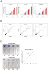
Robustness of the FHAA immunochromatographic test. (A) Six positive samples with different levels of FH-FHAA complexes and two negative controls were tested four times with cassettes from the same batch. The results demonstrate excellent reproducibility of the FHAA ICT. (B) Similarly, 109 samples from our testing cohort were tested with cassettes A, B and C from two different batches and the results also showed excellent reproducibility. (C) Cassettes B from batch 2, generated on June 3, 2023, were used to test the same samples on August 17, 2023, and on June 10, 2024, after being stored in the dark with a desiccant pack at room temperature. They gave essentially identical results, demonstrating excellent stability for at least 1 year. Excel worksheet functions were used for statistical analysis.
Figure 5
Figure 5
Correlation between the intensity of the detection line with free FHAA titers and FH-FHAA complexes. (A) Autoantibody titers in all samples testing positive by ELISA (AU>100) ( Table 1 ) were plotted vs the average of the two measurements of the intensity of the detection band in cassette A. (B) For selected samples, positives by ICT (red dots), the plasma concentration of FH before and after passing through a protein G column was determined to estimate the amount of FH retained in the IgG fraction due to formation of FH FHAA complexes. These FH concentrations were plotted vs the intensity of the detection band in cassette A. Three negative controls were also included in these experiments (green dots). See also Table 5 .
Figure 6
Figure 6
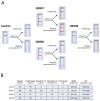
Characterization of IgG FH-FHAA complexes in samples with conflicting ELISA and ICT results. (A) Samples from GN341, GN500, GN198 and from a negative control were passed through a protein G column and the FH concentration was determined in the flow through and the eluate for each sample. The amount of FH retained in the column was determined by subtracting the concentration of FH at the plateau of the elution peak as a more reliable measurement since a variable amount of FH in each sample is lost during the column washes. Cassette B was used to test for the presence of FH-FHAA complexes in the original, the flow through, and the eluted samples. FH-FHAA complexes present in GN341 and GN500 were clearly reduced in the flow through and appear in the eluate, while they were absent in all fractions from GN198 and the control samples. (B) FH concentrations in all samples are shown, together with the ELISA and ICT data. Notice that no FH is detected in the eluate from the GN198 and the control samples. Normal FH range: 90-285µg/ml. NEG, Negative; POS, positive; UND, Undefined.
Figure 7
Figure 7
Characterization of IgM FH-FHAA complexes in samples with conflicting ELISA and ICT results. (A) Results of the analisis by the ICT of plasma samples from cases #18, #34 and #38, from a healthy control (IgG/IgM FHAA negative plasma) and from a strong positive plasma for IgG FHAA that was negative for IgM FHAA (H271B). (B) Plasma from these five samples was passed through an affinity column coated with a goat polyclonal anti-human IgM (Fc fragment) that specifically captures human IgM antibodies and the eluate for each sample tested for the presence of FH by ELISA. The results of these experiments show that while no FH was retained from the healthy control plasma nor the strong positive plasma for IgG FHAA, significant amounts of FH were eluted from the column in the case of the case of the three samples that tested positive for IgM FH-FHAA complexes in the ICT.
Figure 8
Figure 8
Use of ICT to monitor FH-FHAA complexes in patients under immunosuppressive treatment. Serial samples from a patient originally diagnosed with high free FHAA titers, as determined by ELISA, were obtained at different dates (hand written in the cassettes), before, during, and after immunosuppression. For each sample, an ELISA and ICT were performed to determine titers of free FHAA and levels of FH-FHAA complexes.

References

    1. Dragon-Durey MA, Loirat C, Cloarec S, Macher MA, Blouin J, Nivet H, et al. . Anti-factor H autoantibodies associated with atypical hemolytic uremic syndrome. J Am Soc Nephrol. (2005) 16:555–63. doi: 10.1681/ASN.2004050380 - DOI - PubMed
    1. Rodríguez De Córdoba S, Esparza-Gordillo J, Goicoechea De Jorge E, Lopez-Trascasa M, Sánchez-Corral P. The human complement factor H: Functional roles, genetic variations and disease associations. Mol Immunol. (2004) 41:355–67. doi: 10.1016/j.molimm.2004.02.005 - DOI - PubMed
    1. Durey M-AD, Sinha A, Togarsimalemath SK, Bagga A. Anti-complement-factor H-associated glomerulopathies. Nat Rev Nephrol. (2016) 12:563–78. doi: 10.1038/nrneph.2016.99 - DOI - PubMed
    1. Dragon-Durey MA, Sethi SK, Bagga A, Blanc C, Blouin J, Ranchin B, et al. . Clinical features of anti-factor H autoantibody-associated hemolytic uremic syndrome. J Am Soc Nephrol. (2010) 21:2180–7. doi: 10.1681/ASN.2010030315 - DOI - PMC - PubMed
    1. Hofer J, Janecke AR, Zimmerhackl LB, Riedl M, Rosales A, Giner T, et al. . Complement factor H–related protein 1 deficiency and factor H antibodies in pediatric patients with atypical hemolytic uremic syndrome. Clin J Am Soc Nephrol. (2013) 8:407–15. doi: 10.2215/CJN.01260212 - DOI - PMC - PubMed

LinkOut - more resources