

Combination Fractional Carbon Dioxide Laser Treatment and Bone Marrow Mesenchymal Stem Cell Therapy Enhances the Treatment of Skin Photoaging in a Murine Model System
- PMID: 39927128
- PMCID: PMC11803962
- DOI: 10.2147/CCID.S490225
Combination Fractional Carbon Dioxide Laser Treatment and Bone Marrow Mesenchymal Stem Cell Therapy Enhances the Treatment of Skin Photoaging in a Murine Model System
Abstract
Background: Fractional carbon dioxide lasers and bone marrow mesenchymal stem cells (BMSCs) are commonly employed in the treatment of skin photoaging.
Objective: This study was developed to explore the effects of combination carbon dioxide laser treatment and BMSC injection on skin photoaging and the underlying molecular mechanisms.
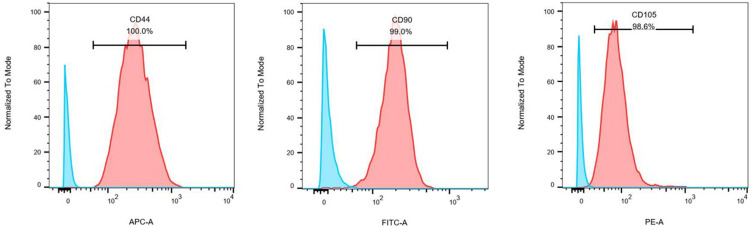
Methods & materials: In total, 24 mice with experimentally photoaged skin were separated into control, carbon dioxide fractional laser treatment, combination therapy, and BMSC injection groups. Samples of dorsal skin from these animals were subjected to hematoxylin and eosin staining or Masson's trichrome staining. In addition, immunohistochemical analyses and real-time polymerase chain reaction analyses were conducted to detect MMP-3 and MMP-9 expression.
Results: After 1 week, both dermal thickness and collagen fiber density were significantly increased in the BMSC and combination treatment groups as compared to the control group (P<0.05), while both of these parameters were significantly increased in all treatment groups after 4 weeks relative to the control group (P<0.05), with the most pronounced effect in the combination therapy group (P<0.05). MMP-3 and MMP-9 mRNA and protein levels in the treatment groups were decreased relative to the control group after 4 weeks.
Conclusion: Combination BMSC and carbon dioxide laser therapy was more effective than either of these therapeutic approaches in isolation as a treatment for photoaged skin. The improvement of effect may be due to the decrease of MMP-3 and MMP-9 expression in combination therapy.
Keywords: bone marrow mesenchymal stem cell; fractional carbon dioxide laser; matrix metalloproteinase; mouse model; photoaging.
© 2025 Li et al.
Conflict of interest statement
The authors report no conflicts of interest in this work.
Figures


References
-
- Fisher G, Kang S, Varani J, et al. Mechanisms of photoaging and chronological skin aging. Arch Dermatol. 2002;138(11):1462–1470. - PubMed
-
- Sun Z, Park S, Hwang E, et al. Salvianolic acid B protects normal human dermal fibroblasts against ultraviolet B irradiation-induced photoaging through mitogen-activated protein kinase and activator protein-1 pathways. Photochemistry and Photobiology. 2015;91(4):879–886. doi:10.1111/php.12427 - DOI - PubMed
LinkOut - more resources
Full Text Sources
Miscellaneous
