

Isolation, characterization and liposome-loaded encapsulation of a novel virulent Salmonella phage vB-SeS-01
- PMID: 39927265
- PMCID: PMC11803447
- DOI: 10.3389/fmicb.2025.1494647
Isolation, characterization and liposome-loaded encapsulation of a novel virulent Salmonella phage vB-SeS-01
Abstract
Introduction: Salmonella is a common foodborne pathogenic bacterium, displaying facultative intracellular parasitic behavior, which can help the escape against antibiotics treatment. Bacteriophages have the potential to control both intracellular and facultative intracellular bacteria and can be developed as antibiotic alternatives.
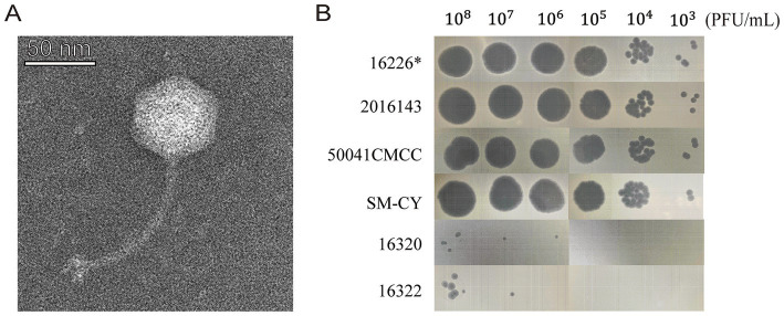
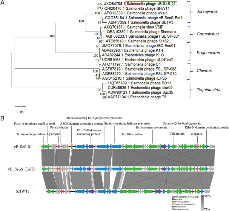
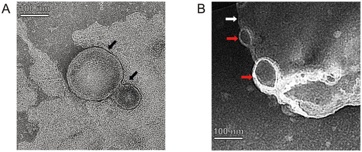
Methods: This study isolated and characterized vB-SeS-01, a novel Guernseyvirinae phage preying on Salmonella enterica, whose genome is closely related to those of phages SHWT1 and vB-SenS-EnJE1. Furthermore, nine phage-carrying liposome formulations were developed by film hydration method and via liposome extruder.
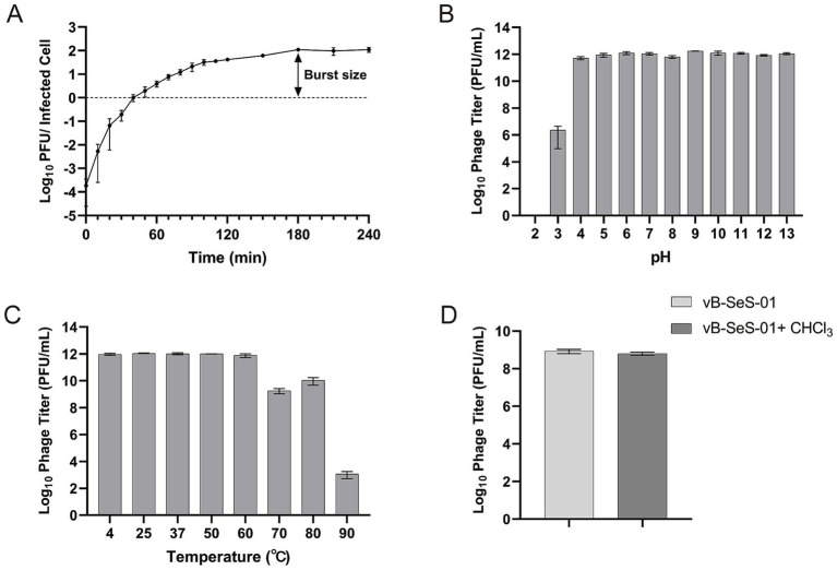
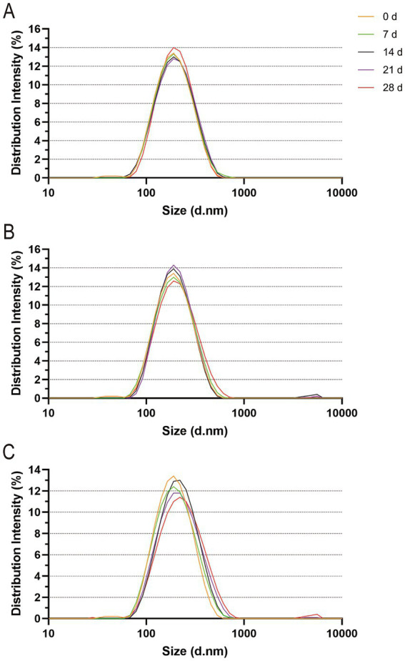
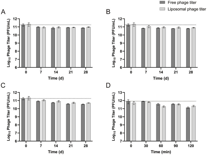
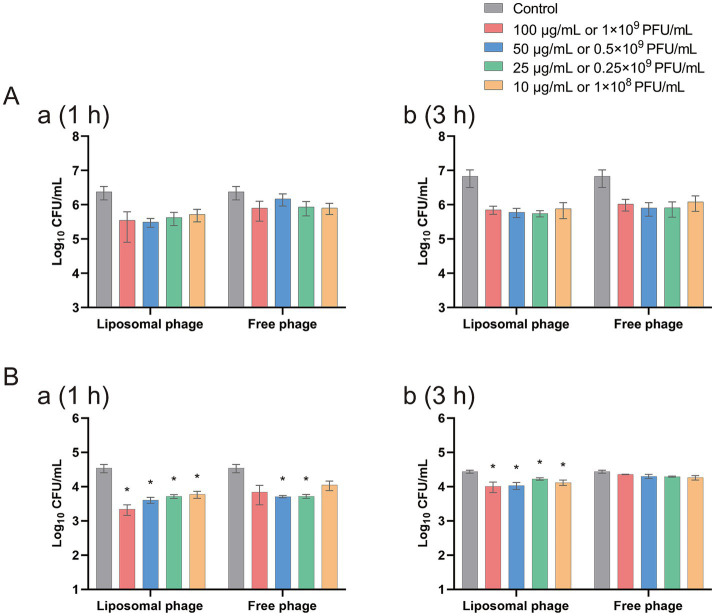
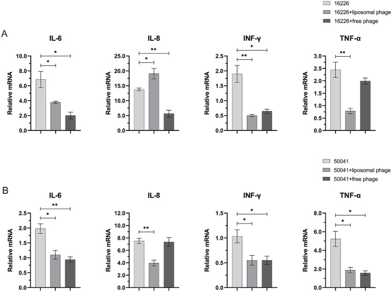
Results and discussion: Phage vB-SeS-01 displays strong lysis ability against 9 out of 24 tested S. enterica strains (including the pathogenic "Sendai" and "Enteritidis" serovars), high replicability with a burst size of 111 ± 15 PFU/ cell and a titre up to 2.1 × 10¹¹ PFU/mL, and broad pH (4.0 ~ 13.0) and temperature (4 ~ 80°C) stabilities. Among the nine vB-SeS-01 liposome-carrying formulations, the one encapsulated with PC:Chol:T80:SA = 9:1:2:0.5 without sonication displayed the optimal features. This formulation carried up to 1011 PFU/mL, with an encapsulation rate of 80%, an average size of 172.8 nm, and a polydispersity index (PDI) of 0.087. It remained stable at 4°C and 23°C for at least 21 days and at 37°C for 7 days. Both vB-SeS-01 and vB-SeS-01-loaded liposomes displayed intracellular antimicrobial effects and could reduce the transcription level of some tested intracellular inflammatory factors caused by the infected S. enterica sv. Sendai 16,226 and Enteritidis 50041CMCC.
Keywords: Guernseyvirinae; Salmonella enterica; intracellular antibacterial effect; liposome; phage.
Copyright © 2025 Luo, Mahillon, Sun, You and Hu.
Conflict of interest statement
The authors declare that the research was conducted in the absence of any commercial or financial relationships that could be construed as a potential conflict of interest.
Figures
Similar articles
-
Efficacy and safety of phage therapy against Salmonella enterica serovars Typhimurium and Enteritidis estimated by using a battery of in vitro tests and the Galleria mellonella animal model.Microbiol Res. 2022 Aug;261:127052. doi: 10.1016/j.micres.2022.127052. Epub 2022 May 4. Microbiol Res. 2022. PMID: 35533436
-
Isolation, characterization, and genome analysis of a broad host range Salmonella phage vB_SenS_TUMS_E4: a candidate bacteriophage for biocontrol.Vet Res Commun. 2023 Sep;47(3):1493-1503. doi: 10.1007/s11259-023-10105-1. Epub 2023 Apr 25. Vet Res Commun. 2023. PMID: 37097546
-
Characterization and genome analysis of a broad host range lytic phage vB_SenS_TUMS_E19 against Salmonella enterica and its efficiency evaluation in the liquid egg.Can J Microbiol. 2024 Sep 1;70(9):358-369. doi: 10.1139/cjm-2024-0013. Epub 2024 Jul 4. Can J Microbiol. 2024. PMID: 38990097
-
Isolation and identification of the broad-spectrum high-efficiency phage vB_SalP_LDW16 and its therapeutic application in chickens.BMC Vet Res. 2022 Nov 3;18(1):386. doi: 10.1186/s12917-022-03490-3. BMC Vet Res. 2022. PMID: 36329508 Free PMC article.
-
Isolation, characterization, therapeutic potency, and genomic analysis of a novel bacteriophage vB_KshKPC-M against carbapenemase-producing Klebsiella pneumoniae strains (CRKP) isolated from Ventilator-associated pneumoniae (VAP) infection of COVID-19 patients.Ann Clin Microbiol Antimicrob. 2023 Feb 24;22(1):18. doi: 10.1186/s12941-023-00567-1. Ann Clin Microbiol Antimicrob. 2023. PMID: 36829156 Free PMC article.
References
LinkOut - more resources
Full Text Sources