

Ablation of Htra1 leads to sub-RPE deposits and photoreceptor abnormalities
- PMID: 39927462
- PMCID: PMC11948579
- DOI: 10.1172/jci.insight.178827
Ablation of Htra1 leads to sub-RPE deposits and photoreceptor abnormalities
Abstract
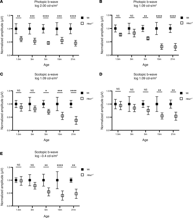
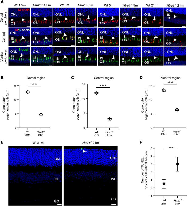
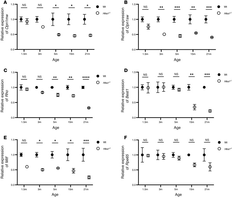
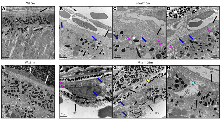
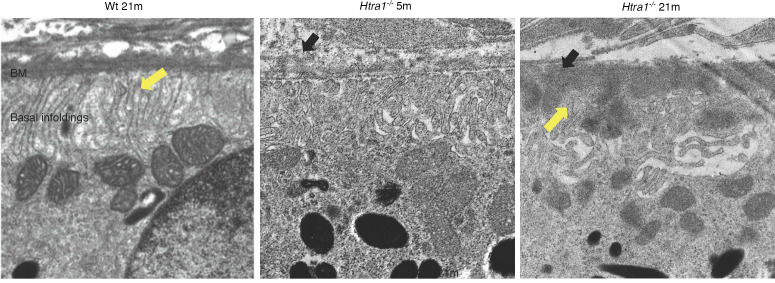
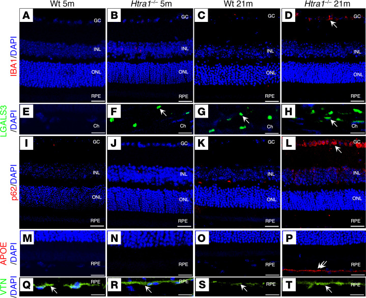
The high-temperature requirement A1 (HTRA1), a serine protease, has been demonstrated to play a pivotal role in the extracellular matrix (ECM) and has been reported to be associated with the pathogenesis of age-related macular degeneration (AMD). To delineate its role in the retina, the phenotype of homozygous Htra1-KO (Htra1-/-) mice was characterized to examine the effect of Htra1 loss on the retina and retinal pigment epithelium (RPE) with age. The ablation of Htra1 led to a significant reduction in rod and cone photoreceptor function, primary cone abnormalities followed by rods, and atrophy in the RPE compared with WT mice. Ultrastructural analysis of Htra1-/- mice revealed RPE and Bruch's membrane (BM) abnormalities, including the presence of sub-RPE deposits at 5 months (m) that progressed with age accompanied by increased severity of pathology. Htra1-/- mice also displayed alterations in key markers for inflammation, autophagy, and lipid metabolism in the retina. These results highlight the crucial role of HTRA1 in the retina and RPE. Furthermore, this study allows for the Htra1-/- mouse model to be utilized for deciphering mechanisms that lead to sub-RPE deposit phenotypes including AMD.
Keywords: Genetics; Mouse models; Neurodegeneration; Ophthalmology; Retinopathy.
Figures
Similar articles
-
Chromosome 10q26-driven age-related macular degeneration is associated with reduced levels of HTRA1 in human retinal pigment epithelium.Proc Natl Acad Sci U S A. 2021 Jul 27;118(30):e2103617118. doi: 10.1073/pnas.2103617118. Proc Natl Acad Sci U S A. 2021. PMID: 34301870 Free PMC article.
-
Altered Elastin Turnover, Immune Response, and Age-Related Retinal Thinning in a Transgenic Mouse Model With RPE-Specific HTRA1 Overexpression.Invest Ophthalmol Vis Sci. 2024 Jul 1;65(8):34. doi: 10.1167/iovs.65.8.34. Invest Ophthalmol Vis Sci. 2024. PMID: 39028977 Free PMC article.
-
High-Temperature Requirement A 1 Causes Photoreceptor Cell Death in Zebrafish Disease Models.Am J Pathol. 2018 Dec;188(12):2729-2744. doi: 10.1016/j.ajpath.2018.08.012. Epub 2018 Sep 29. Am J Pathol. 2018. PMID: 30273602
-
Secretory proteostasis of the retinal pigmented epithelium: Impairment links to age-related macular degeneration.Prog Retin Eye Res. 2020 Nov;79:100859. doi: 10.1016/j.preteyeres.2020.100859. Epub 2020 Apr 9. Prog Retin Eye Res. 2020. PMID: 32278708 Review.
-
Cellular Senescence: An Emerging Player in the Pathogenesis of AMD.Adv Exp Med Biol. 2025;1468:33-37. doi: 10.1007/978-3-031-76550-6_6. Adv Exp Med Biol. 2025. PMID: 39930169 Review.
Cited by
-
Retinal Autophagy for Sustaining Retinal Integrity as a Proof of Concept for Age-Related Macular Degeneration.Int J Mol Sci. 2025 Jun 16;26(12):5773. doi: 10.3390/ijms26125773. Int J Mol Sci. 2025. PMID: 40565235 Free PMC article. Review.
References
MeSH terms
Substances
Grants and funding
LinkOut - more resources
Full Text Sources
Medical
Research Materials
