. 2025 Feb 10;10(3):e186096.
doi: 10.1172/jci.insight.186096.

Prenatal alcohol exposure is associated with altered feto-placental blood flow and sex-specific placental changes

Affiliations

Prenatal alcohol exposure is associated with altered feto-placental blood flow and sex-specific placental changes

Sarah E Steane et al. JCI Insight. .

Abstract

BACKGROUNDPrenatal alcohol exposure (PAE) around conception in preclinical models results in placental insufficiency, likely contributing to offspring abnormalities. Altered placental DNA methylation (DNAm) and gene expression suggest epigenetic mechanisms, perhaps involving impacts on methyl donor levels. PAE around conception in women is common but placental effects are rarely examined. This cohort study investigated associations between PAE around conception and intake/plasma measures of the methyl donors folate and choline, feto-placental blood flow, and placental growth measures, gene expression, and DNAm.METHODSPregnant participants delivered at Mater Mothers' Hospital, Brisbane, Queensland, Australia (n = 411). Dietary intake of choline and folate were calculated and plasma concentrations measured using mass spectrometry (MS) and clinical immunoanalyzer, respectively. Cerebroplacental ratio (CPR) was calculated using Doppler measurements. Placentas were weighed/measured at delivery and samples used to quantify methyl donors (MS), global DNAm (ELISA), and gene expression (quantitative PCR). Data were compared between control/abstinent and PAE groups, by fetal sex.RESULTSA CPR <5th-centile, indicating fetal brain sparing because of placental insufficiency, was found in 2% of controls and 18% of the PAE group, mostly male fetuses (63%). Compared with controls, male PAE placentas had reduced mean thickness and placental growth factor mRNA and DNAm, whereas female PAE placentas had increased S-adenosylmethionine and a trend toward increased DNAm.CONCLUSIONPAE around conception is associated with reduced CPR and altered placental growth measures, particularly in males, with potential implications for future health.FUNDINGNational Health and Medical Research Council (APP1191217) and Mary McConnel Career Boost Program for Women in Paediatric Research (WIS132020).

Keywords: Development; Growth factors; Molecular biology; Obstetrics/gynecology; Reproductive biology.


Conflict of interest statement

Conflict of interest: The authors have declared that no conflict of interest exists.

Figures

Figure 1
Figure 1. Flow diagram of study.
Number of participants enrolled, withdrawals/exclusions, and final number included. Sample sizes for participant data/samples are shown with details of analyses. 5MTHF, 5-methyltetrahydrofolate; SAM, S-adenosylmethionine; PlGF, placental growth factor; VEGFA, vascular endothelial growth factor; FLT1, VEGF receptor 1; KDR, VEGF receptor 2; DNMT, DNA methyltransferase; MTOR, mechanistic target of rapamycin; RFC, reduced folate carrier.
Figure 2
Figure 2. Doppler ultrasound measurements across gestation in a general pregnancy cohort.
Measurements of umbilical artery pulsatility index (UAPI) and fetal middle cerebral artery pulsatility index (MCAPI) and cerebroplacental ratio across gestation (24, 28, and 36 weeks) in male (AC) and female (DF) fetuses and at 36 weeks’ gestation in male and female fetuses (GI). Women were grouped by alcohol consumption into abstinent (con, green: male n = 24, female n = 21), alcohol preconception only (PC, light blue: male n = 33, female n = 38), or alcohol preconception and during pregnancy (PCP, dark blue: male n = 27, female n = 21). Data expressed as mean ± SEM. Measurements compared between groups over time (AF), using a mixed effects model, and at 36 weeks (GI), using 2-way ANOVA and Tukey’s post hoc test. Residuals were normally distributed. P < 0.05 was considered statistically significant. Fetuses with CPR <5th-centile indicated by gray data points (I). Complete data at 36 weeks. Missing UAPI and/or MCAPI for males at 24 and 28 weeks con (n = 2), PC (n = 1), and 28 weeks PCP (n = 1) and for females at 24 and 28 weeks con (n = 2), 24 weeks PC (n = 4), and 28 weeks PC (n = 1).
Figure 3
Figure 3. Placental expression of growth factors by alcohol and sex.
Expression of placental growth factor (PlGF) (AD), VEGF receptor 1 (FLT1) (EH), and PlGF/FLT1 (IL). Placentas collected from abstinent women (con: male n = 40, female con n = 31) and women reporting prenatal alcohol exposure (PAE) (male n = 95, female n = 112). The PAE group was further divided by timing into preconception only (PC: male n = 51, female n = 73) or preconception and during pregnancy (PCP: male n = 44, female n = 39). Gene expression normalized to β-actin (ACTB), and fold-change expressed relative to male con group. Data expressed as mean ± SEM. Con and PAE groups compared using unpaired t test/1-way ANOVA where residuals were normally distributed or where transformation normalized distribution. Otherwise, Mann-Whitney/Kruskal-Wallis tests were used (E, F, H, and J). P < 0.05 was considered statistically significant, and P = 0.05–0.1 was considered a trend. FLT1 was below detection limit for 1 male control, as was PlGF for 1 female PC.
Figure 4
Figure 4. Placental expression of DNMTs by alcohol and sex.
Expression of DNA methyltransferases DNMT1 (AD), DNMT3a (EH), and DNMT3b (IL). Placentas collected from abstinent women (con: male n = 40, female n = 31) and women reporting prenatal alcohol exposure (PAE) (male n = 95, female n = 112). The PAE group was also divided by timing into preconception only (PC: male n = 51, female n = 73) or preconception and during pregnancy (PCP: male n = 44, female n = 39). Gene expression normalized to ACTB, and fold-change expressed relative to the male con group. Data expressed as mean ± SEM. Con and PAE groups compared using unpaired t test/1-way ANOVA where residuals were normally distributed or where transformation normalized distribution (I and J). Otherwise, Mann-Whitney/Kruskal-Wallis tests were used. P < 0.05 was considered statistically significant, and P = 0.05–0.1 was considered a trend. *P < 0.05 for Tukey’s multiple comparisons. DNMT3a was below detection limit in 1 female PC placenta, as was DNMT3b in 2 female PC placentas.
Figure 5
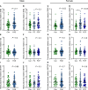
Figure 5. Relative levels of methyl donors in the placenta by alcohol and sex.
Relative levels of S-adenosylmethionine (SAM) (AD), methionine (met) (EH), and 5-methyltetrahydrofolate (5MTHF) (IL). Placentas collected from abstinent women (con: male n = 40, female n = 31) or women reporting prenatal alcohol exposure (PAE) (male n = 95, female n = 111). The PAE group was further divided by timing of exposure into preconception only (PC: male n = 51, female n = 72) or preconception and during pregnancy (PCP: male n = 44, female n = 39). The ratio of methyl donor/internal standard was calculated and mean fold-change ± SEM expressed relative to the male con group. Expression differences across groups compared using unpaired t test/1-way ANOVA where residuals were normally distributed or where transformation normalized distribution. Otherwise, Mann-Whitney/Kruskal-Wallis tests were used (G and H). P < 0.05 was considered statistically significant, and P = 0.05–0.1 was considered a trend. *P < 0.05 for Tukey’s multiple comparisons. One female PC sample failed mass spectrometry.
Figure 6
Figure 6. Placental expression of nutrient regulators by alcohol and sex.
Expression of mechanistic target of rapamycin (MTOR) (AD) and reduced folate carrier (RFC) (EH). Placentas collected from abstinent women (con: male n = 40, female con n = 31) or women with prenatal alcohol exposure (PAE) (male n = 95, female n = 112). The PAE group was also divided by timing of exposure into preconception only (PC: male n = 51, female n = 73) or preconception and during pregnancy (PCP: male n = 44, female n = 39). Gene expression normalized to ACTB, and fold-change expressed relative to male con group. Data expressed as mean ± SEM. Con and PAE groups compared using unpaired t test/1-way ANOVA where residuals were normally distributed or where transformation normalized residuals. Otherwise, Mann-Whitney/Kruskal-Wallis tests were used (CH). P < 0.05 was considered statistically significant, and P = 0.05–0.1 was considered a trend. *P < 0.05 for Tukey’s multiple comparisons test. MTOR was below detection limit in female con (n = 1), PC (n = 2), and PCP (n = 1), as was RFC in female PC (n = 2).

References

    1. Kesmodel U, et al. Moderate alcohol intake in pregnancy and the risk of spontaneous abortion. Alcohol Alcohol. 2002;37(1):87–92. doi: 10.1093/alcalc/37.1.87. - DOI - PubMed
    1. Patra J, et al. Dose-response relationship between alcohol consumption before and during pregnancy and the risks of low birthweight, preterm birth and small for gestational age (SGA)-a systematic review and meta-analyses. BJOG. 2011;118(12):1411–1421. doi: 10.1111/j.1471-0528.2011.03050.x. - DOI - PMC - PubMed
    1. Aliyu MH, et al. Alcohol consumption during pregnancy and the risk of early stillbirth among singletons. Alcohol. 2008;42(5):369–374. doi: 10.1016/j.alcohol.2008.04.003. - DOI - PubMed
    1. Popova, S , et al. Fetal alcohol spectrum disorders. Nat Rev Dis Primers. 2023;9:11. doi: 10.1038/s41572-023-00420-x. - DOI - PubMed
    1. O’Keeffe LM, et al. Prevalence and predictors of alcohol use during pregnancy: findings from international multicentre cohort studies. BMJ Open. 2015;5(7):e006323. doi: 10.1136/bmjopen-2014-006323. - DOI - PMC - PubMed