. 2025 Apr;12(13):e2417449.
doi: 10.1002/advs.202417449. Epub 2025 Feb 10.

Hybrid Electro-optical Stimulation Improves Ischemic Brain Damage by Augmenting the Glymphatic System

Affiliations

Hybrid Electro-optical Stimulation Improves Ischemic Brain Damage by Augmenting the Glymphatic System

Min Jae Kim et al. Adv Sci (Weinh). 2025 Apr.

Abstract

Ischemic brain injury not only results in significant neurological, motor, and cognitive impairment but also contributes to the accumulation of toxic solutes and proinflammatory cytokines in the infarction region, exacerbating ischemic brain damage. The glymphatic system, which is crucial for brain waste clearance and homeostasis, is impaired by ischemic injury, highlighting the importance of developing therapeutic strategies for poststroke complications. Herein, a novel hybrid electro-optical stimulation device is proposed that integrates near-infrared micro-light-emitting diode with transparent microneedles, enabling efficient noninvasive stimulation of the cortical area for ischemic stroke treatment. This study investigates whether this hybrid electro-optical stimulation enhances the glymphatic system function and ameliorates ischemic brain injury in the middle cerebral artery occlusion and reperfusion (MCAO/R) mice model. The results demonstrate that hybrid stimulation improves the neurological, motor, and cognitive functions and reduces brain atrophy following MCAO/R. Moreover, hybrid stimulation restores impaired glymphatic system function by modulation of aquaporin-4 (AQP4) polarization and alleviates the accumulation of proinflammatory cytokines such as IL-1β. Notably, AQP4 inhibition partly reverses the improved functional outcomes of hybrid stimulation. The findings suggest that targeting glymphatic drainage using hybrid electro-optical stimulation is a promising therapeutic approach for treating ischemic brain injury.

Keywords: aquaporin‐4 polarization; cerebrospinal fluid; glymphatic system; hybrid electro‐optical stimulation; ischemic stroke.


Conflict of interest statement

The authors declare no conflict of interest.

Figures
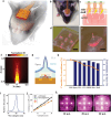
Figure 1
Figure 1
Concept and optical characterization of hybrid electro‐optical stimulator based on a transparent conductive microneedle array (MNA) for stroke rehabilitation in a mouse model. a) Illustration and b) photograph of the device and animal for hybrid electro‐optical stimulation. c) Cross‐sectional presentation of hybrid electro‐optical stimulation. d) Photograph of light emitted through a PLA MNA and e) a magnified view of a single needle (630 nm µLEDs were used here for visual demonstration of optical emission). Scale bar: 2 mm (d) and 0.5 mm (e). f) Monte Carlo simulation of the spatial distribution of normalized light intensity (Φ) from a µLED underneath PLA microneedle. g) Cross‐sectional structure of a PLA microneedle consisting of a µLED pocket, conductive ITO layer, and insulating PDMS layer. h) Optical transmittance (850 nm) of each layer between µLED and cortex, and cumulative transmittance from µLED to cortex. i) Emission spectrum of 850 nm µLED. j) Irradiance of light emitted from a µLED and four PLA MLA devices. k) Photographs showing increasing light intensity with varying driving currents from F, 10 to 30 mA (750 nm IR filter was used to visualize the 850 nm light). Abbreviations: Polydimethylsiloxane (PDMS), polylactide (PLA), and indium tin oxide (ITO).
Figure 2
Figure 2
Fabrication and electromechanical characterization of the hybrid electro‐optical stimulator. a) Exploded structure of the stimulator. b) Simplified fabrication process of PLA MNA. Master mold of acupuncture needles (i) is double‐casted (ii) by PLA to form MNA with underside µLED pockets (iii), followed by ITO coating (iv) and assembly with µLED‐mounted FPCB cable and PDMS insulation (v). c,d) Comparison of master microneedle and PLA microneedle, with quantitative analysis of their geometric parameters. Scale bar: 0.2 mm. e) Lateral bending force measured from PLA MNA with (red) and without meniscus‐shaped base (blue). f) Three‐dimensional mouse brain model with skull, scalp, and electrode placement for simulation of electrical stimulation from the device. Simulated distribution of g) electric field and h) current density at the cortex surface. Abbreviations: height (H), total height (TH), width (W), light‐emitting diode (LED), microneedle array (MNA), polydimethylsiloxane (PDMS), polylactide (PLA), and indium tin oxide (ITO).
Figure 3
Figure 3
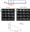
Effects of hybrid electro‐optical stimulation on infarction size following MCAO/R over time. a) Schematic timeline showing the experimental setup and stimulation periods for MCAO/R and hybrid electro‐optical stimulation. MRI scans were performed before stimulation (on day 2) and on days 4, 6, and 16 after MCAO/R. b) Representative T2‐weighted MRI images showing infarct areas in stimulated (hybrid) and non‐stimulated (MCAO/R) groups. c) Quantification of infarction size (mm2) over time, demonstrating a significant reduction in infarct volume in the hybrid stimulation group compared to that in the MCAO/R group, particularly on days 4 and 6. All data are represented as mean ± SEM (n = 4 per group). Statistical significance was determined using unpaired, two‐tailed Student's t‐tests. *P < 0.05, **P < 0.01, versus the MCAO/R group. Abbreviations: middle cerebral artery occlusion/reperfusion (MCAO/R).
Figure 4
Figure 4
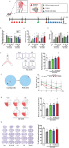
Hybrid electro‐optical stimulation improves functional recovery and brain atrophy after ischemic injury. a) Experimental design. The mice underwent behavior tests to evaluate the recovery of b) neurological deficits and motor function using c) rotarod and d) wire grip tests at 7 and 14 d after ischemic injury (N = 8 per group). e) (Left panel) The mice freely explored the Y‐maze for 8 min while being recorded for (middle panel) tracking movement. e) (Right panel) Quantification of spontaneous alternation rate (N = 8 per group). f) (Left panel) Spatial learning and memory were evaluated using Morris water maze test. (Right panel) During learning trials, the mice were placed in a water‐filled pool with a hidden platform for learning trial tests and measured escape latency. On probe trial day, mice were conducted to evaluate the time spent in the target quadrant without the platform for 90 s. g (Left panel) During this trial, the mice were recorded for tracking movement. (Right panel) Quantification of the percentage of spent time in the target quadrant (N = 8 per group). h) Twenty‐five days after MCAO/R, the obtained brain slices were stained with 0.1% cresyl violet, and brain atrophy was quantified (N = 5 per group). All data are represented as mean ± SEM. Statistical significance was determined using one‐way ANOVA with Tukey's post‐hoc test. *P < 0.05 and ***P < 0.001, versus the control group. #P < 0.05, ##P < 0.01, and ###P < 0.001, versus the MCAO/R group. $P < 0.05 versus the ES group. &&P < 0.01 versus the OS group. Abbreviations: neurological severity score (NSS), control (Con), middle cerebral artery occlusion/reperfusion (MCAO/R), electrical stimulation (ES), and optical stimulation (OS).
Figure 5
Figure 5
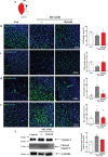
Hybrid electro‐optical stimulation enhances neuroprotective effects on the ischemic brain. a) Illustration of the ROI for observing immunostained markers in the peri‐infarct regions 7 d after MCAO/R. Representative images and quantification of b) NeuN (Neuronal cell), c) CD31 (endothelial cell), d) GFAP (astrocyte), and e) Iba‐1 (microglia). N = 9 images for each group. f) Representative western blot images showing caspase‐3 (30 kDa), cleaved caspase‐3 (17 kDa), and α‐tubulin (50 kDa, loading control), as well as the quantification of caspase‐3 and cleaved caspase‐3 expression relative to α‐tubulin (N = 4 for each group). All data are represented as mean ± SEM. Statistical significance was determined using one‐way ANOVA with Tukey's post‐hoc test. *P < 0.05, **P < 0.01, and ***P < 0.001, versus the control group. #P < 0.05, ##P < 0.01, and ###P < 0.001, versus the MCAO/R group. Abbreviations: control (Con), middle cerebral artery occlusion/reperfusion (MCAO/R), region of interest (ROI), neuronal cell (NeuN), cluster differentiation 31 (CD31), glial fibrillary acidic protein (GFAP), ionized calcium‐binding adapter molecule‐1 (Iba‐1), and 4′,6‐diamidino‐2‐phenylindole (DAPI).
Figure 6
Figure 6
Hybrid electro‐optical stimulation restores glymphatic function in the ischemic brain. a) (Left panel) Experimental design. Mice were intraperitoneally administered TGN‐020 (200 mg kg−1) 10 min after MCAO/R for inhibition of AQP4. (Right panel) The illustration shows that a fluorescence tracer was injected into the cisterna magna or brain parenchyma to assess glymphatic function 7 d after MCAO/R. Seven coronal brain sections at 1.1, 0.5, 0.14, −0.46, −0.94, −1.34, and −1.94 from bregma per animal were quantified. b) Representative images show the fluorescence tracer influx and average quantification of fluorescence intensity of CSF tracer efflux into the brain parenchyma (N = 21 brain slices per group). c) Representative images indicate the fluorescence tracer of CSF clearance and quantification of percentage area of the accumulated CSF tracer per brain slice (N = 21 brain slices per group). d) Representative western blot images showing IL‐1β (32 kDa) and α‐tubulin (50 kDa, loading control), along with the quantification of IL‐1β expression relative to α‐tubulin (N = 4 for each group). e) Representative images show proinflammatory cytokine, IL‐1β staining in the peri‐infarct area, and quantification of fluorescence intensity (N = 8 images for each group). All data are represented as mean ± SEM. Statistical significance was determined using one‐way ANOVA with Tukey's post‐hoc test. **P < 0.01 and ***P < 0.001 versus the control group. #P < 0.05 and ###P < 0.001, versus the MCAO/R group. $P < 0.05, $$P < 0.01, and $$$P < 0.001, versus the hybrid group. Abbreviations: cerebrospinal fluid (CSF), control (Con), middle cerebral artery occlusion/reperfusion (MCAO/R), TGN‐020 (TGN), Interleukin‐1 beta (IL‐1β), and 4′,6‐diamidino‐2‐phenylindole (DAPI).
Figure 7
Figure 7
Hybrid electro‐optical stimulation recovers AQP4 polarization in the ischemic brain. a) Representative images of AQP4/GFAP immuno‐staining in the peri‐infarct cortex 7 d after MCAO/R. White arrows of 40× magnification images represent the localized AQP4 on astrocytic endfeet. b) Representative images show AQP4/CD31 in the peri‐infarct cortex 7 d after MCAO/R and 40× magnification images represent AQP4 coverage of vessels. c–e) Quantification of the AQP4‐positive area (N = 19 images per group), perivascular AQP4 polarization (N = 30 vessels per group), and AQP4 coverage of CD31 (N = 9 images per group). All data are represented as mean ± SEM. Statistical significance was determined using one‐way ANOVA with Tukey's post‐hoc test. **P < 0.01 and ***P < 0.001 versus the control group. #P < 0.05 and ##P < 0.01, versus the MCAO/R group. $P < 0.05 and $$$P < 0.001, versus the Hybrid group. Abbreviations: control (Con), middle cerebral artery occlusion/reperfusion (MCAO/R), TGN‐020 (TGN), Aquaporin‐4 (AQP4), glial fibrillary acidic protein (GFAP), and cluster differentiation 31 (CD31).
Figure 8
Figure 8
AQP4 inhibition reverses the beneficial effects of hybrid electro‐optical stimulation on ischemic injury. a) Experimental design. Mice were intraperitoneally administered TGN‐020 (200 mg kg−1) 10 min after MCAO/R and treated with hybrid electro‐optical stimulation for 5 d. The mice underwent behavior tests to evaluate the recovery of b) neurological deficits and motor function using c) rotarod and d) wire grip tests at 7 and 14 d after ischemic injury. e) Mice freely explored the Y‐maze for 8 min and quantified the spontaneous alternation rate. Spatial learning and memory were evaluated using the Morris water maze test. f) Mice were placed in a water‐filled pool with a hidden platform for learning trial tests and measured escape latency during learning trials. g) On day 7, a probe trial test was conducted to evaluate the time the mice spent in the target quadrant without the platform for 90 s. All data are represented as mean ± SEM. N = 10 per group. Statistical significance was determined using one‐way ANOVA with Tukey's post‐hoc test. *P < 0.05, **P < 0.01, and ***P < 0.001, versus the MCAO/R group. #P < 0.05, #P < 0.05, and ###P < 0.001, versus the MCAO/R group. Abbreviations: middle cerebral artery occlusion/reperfusion (MCAO/R), and TGN‐020 (TGN).

References

    1. Stankowski J. N., Gupta R., Antioxid. Redox Signaling 2011, 14, 1841. - PMC - PubMed
    1. Martin S. S., Aday A. W., Almarzooq Z. I., Anderson C. A. M., Arora P., Avery C. L., Baker‐Smith C. M., Barone Gibbs B., Beaton A. Z., Boehme A. K., Commodore‐Mensah Y., Currie M. E., Elkind M. S. V., Evenson K. R., Generoso G., Heard D. G., Hiremath S., Johansen M. C., Kalani R., Kazi D. S., Ko D., Liu J., Magnani J. W., Michos E. D., Mussolino M. E., Navaneethan S. D., Parikh N. I., Perman S. M., Poudel R., Rezk‐Hanna M., et al., Circulation 2024, 149, e347. - PubMed
    1. a) Anrather J., Iadecola C., Neurotherapeutics 2016, 13, 661; - PMC - PubMed
    2. b) Zbesko J. C., Nguyen T. V., Yang T., Frye J. B., Hussain O., Hayes M., Chung A., Day W. A., Stepanovic K., Krumberger M., Mona J., Longo F. M., Doyle K. P., Neurobiol. Dis. 2018, 112, 63. - PMC - PubMed
    1. Esposito E., Ahn B. J., Shi J., Nakamura Y., Park J. H., Mandeville E. T., Yu Z., Chan S. J., Desai R., Hayakawa A., Ji X., Lo E. H., Hayakawa K., Nat. Commun. 2019, 10, 5306. - PMC - PubMed
    1. Back D. B., Kwon K. J., Choi D. H., Shin C. Y., Lee J., Han S. H., Kim H. Y., J. Neuroinflammation 2017, 14, 216. - PMC - PubMed

LinkOut - more resources