. 2025 Feb 10;82(1):74.
doi: 10.1007/s00018-025-05606-y.

SLPI controls neutrophil migration abilities and impacts neutrophil skin infiltration in experimental psoriasis

Affiliations

SLPI controls neutrophil migration abilities and impacts neutrophil skin infiltration in experimental psoriasis

Patrycja Kwiecinska et al. Cell Mol Life Sci. .

Abstract

Skin infiltration by neutrophils is a hallmark of the chronic inflammatory skin disease psoriasis, yet the mechanisms underlying neutrophil recruitment and positioning in chronically inflamed skin remain poorly understood. In this study, we demonstrate the significant impact of a total genetic deficiency of secretory leukocyte protease inhibitor (SLPI) on neutrophil migration in mouse skin. Without SLPI, neutrophils displayed an unconventional migratory pattern, characterized by altered interactions with vessel walls and reduced efficiency in extravasating from blood vessels into skin tissue during the early stages of experimental psoriasis. This was associated with changes in tissue motility, positioning neutrophils farther from the skin entry vessels and closer to the skin surface. Neutrophil diapedesis was partially dependent on SLPI within the neutrophils themselves. The impact of SLPI on neutrophil movement was further supported by the increased migration of human neutrophils in the presence of neutrophil-penetrant recombinant SLPI. Additionally, our data suggest that neutrophils with varying capacities for vessel wall interaction are released from the bone marrow into circulation in an SLPI-dependent manner. These findings establish a role for SLPI in regulating the spatiotemporal infiltration of neutrophils into the skin in psoriasis, highlighting its relevance to psoriasis pathophysiology.

Keywords: Chronic inflammation; Dermis; Inhibitors of serine proteases; Innate immunity; Intravital microscopy; Neutrophil elastase.


Conflict of interest statement

Declarations. Ethical approval: All animal procedures and experiments were performed in accordance with national and European legislation, following approval by the 2nd Local Institutional Animal Care and Use Committee (IACUC) in Krakow (# 298/2017 and 61/2023). All human studies were performed in accordance with guidelines established by the Jagiellonian University Institutional Bioethics Committee under approved protocols (#87/B/2014; 1072.6120.30.2020) and adhered to the Declaration of Helsinki. Human blood was collected from healthy individuals who were fully informed and had consented. Competing interests: The authors have no relevant financial or non-financial interests to disclose.

Figures

Fig. 1
Fig. 1
SLPI KO and WT mice share neutrophil development patterns. Linage-negative cells from the bone marrow of the indicated mice were subjected to staining for c-kit and Ly6G followed by flow cytometry analysis. (a) Representative flow cytometry plot from WT mice showing the gating strategy for immature and mature neutrophils. (b) Number of neutrophils at different stages of development. Data are shown as the mean ± SD. Dots represent individual mice. MB = myeloblasts, PM = promyelocytes, MC = myelocytes, MM = metamyelocytes, PMN = mature neutrophils
Fig. 2
Fig. 2
SLPI influences the kinetics of neutrophil infiltration into the skin in psoriasis-like dermatitis. The indicated mice were subjected to IMQ treatment. (a) Bone marrow and blood were harvested and analyzed by flow cytometry. Data are presented as mean ± SEM, with 3–6 mice per group. A mixed model ANOVA with Greenhouse-Geisser correction followed by Šídák’s multiple comparison test revealed no statistically significant differences between WT and SLPI KO mice. (b) Skin was harvested and analyzed by flow cytometry. Total leukocytes were detected using anti-CD45 mAbs, and neutrophils were identified using anti-Ly6G and CD11b mAbs. Data are presented as mean ± SEM, with n = 6–8 mice per group. Statistical significance (*p < 0.05) was determined using a mixed model ANOVA with Greenhouse-Geisser correction, followed by Šídák’s multiple comparison test. (c) Representative flow cytometry plots (left panel), the number of skin-infiltrating neutrophils (middle panel), and the percentage of skin-infiltrating neutrophils (right panel) at day 2 of IMQ treatment are shown. Dots represent individual mice; bars indicate the mean ± SD, n = 6–8 mice per group. *p < 0.05, **p < 0.01, by Student’s t-test
Fig. 3
Fig. 3
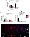
IVM visualizing psoriatic skin vasculature reveals differences in the number of vessel-interacting neutrophils between SLPI KO mice and their WT counterparts. The indicated mice were subjected to IMQ treatment for 2 days. Neutrophils were fluorescently labeled intravenously with Alexa Fluor 647-conjugated anti-mouse Ly6G 24 h before imaging, and with eFluor 450-conjugated anti-mouse Ly6G mAbs together with CD49b mAbs (to detect platelets) just before 4-hr IVM imaging. At least 2 different fields of view per mouse were analyzed. N = 3 mice per group. (a) Ly6G+ cells were counted in circulation (circulating neutrophils in the process of diapedesis) and also outside, but nearby, blood vessels (extravasated neutrophils). Data are presented as mean ± SD. *p < 0.05, ****p < 0.0001 by Student’s t-test. “nd"= not detected. (b) Neutrophils labeled 24 h before imaging (group 1), just before 4-hr imaging (group 2), or labeled by both anti-Ly6G Abs (group 3) are shown. Data are presented as mean ± SD. **p < 0.01, ***p < 0.001, ****p < 0.0001, by one-way ANOVA (Bonferroni post-hoc). “nd” indicates cells were not detected in this area. (c) Representative images of mouse ear skin depicting differences in the number and location of neutrophils in WT and SLPI KO mice. The blood vessel is marked with a white dotted line, exemplary neutrophils (purple) are marked with white arrows, platelets (red) that were used to indicate blood flow are marked with yellow arrowheads. Autofluorescent hair follicles are marked with asterisks. Scale bar = 50 μm
Fig. 4
Fig. 4
SLPI deficiency alters the neutrophil migration pattern in their interaction with vessels at the early stages of experimental psoriasis. The indicated mice were subjected to IMQ treatment for 2 days. Neutrophils were fluorescently labeled intravenously with anti-Ly6G mAbs, and platelets with anti-CD49b mAbs, followed by IVM imaging. One 10-min. movie was analyzed for each mouse. N = 3 mice per group. (a) Time in seconds needed for neutrophils to travel a distance of 50 μm inside the ear skin blood vessel. Data are presented as mean ± SD, ***p < 0.001 by Student’s t-test. (b) Representative images of mouse ear skin capturing the motion of neutrophils within the blood vessels. Points indicate the current location of a neutrophil in a chosen timeframe, while white lines depict the path traveled. Exemplary neutrophil trajectories are shown, with the red arrow indicating the current cell position. (c) Percentage of neutrophils executing “jumps” within the blood vessels. Data are presented as mean ± SD. “nd” =not detected
Fig. 5
Fig. 5
In SLPI-deficient mice, there are more extravasated neutrophils far from the vessel compared to WT mice. The indicated mice were subjected to IMQ treatment for 2 days. Neutrophils were fluorescently labeled intravenously with anti-Ly6G mAbs, and platelets with anti-CD49b mAbs, followed by IVM imaging. (a) Schematic of the counting strategy: A series of optical cross-sections (z-stacks) were made through the ear skin. An arbitrary mask was applied to estimate the number of neutrophils at three different levels above the blood vessel, where level 1 is the closest to the vessel and level 3 is the closest to the skin surface. (b) Exemplary images of z-stacks made through the skin of SLPI KO mice and their WT counterparts. A blood vessel (marked with a yellow arrowhead) is identified by platelet flow (red). Neutrophils are stained in violet. (c) Extravasated neutrophils were counted in arbitrary compartments above the blood vessel. Data are present as mean ± SD * p < 0.05, ** p < 0.01, *** p < 0.001, **** p < 0. 0001 by Student’s t-test. A minimum of 3 intravital movies were analyzed per mouse, n = 2 mice per group. (d) Cell movement: absolute path length of neutrophils at different levels of psoriatic ear skin tissue above the blood vessel from which they extravasated in SLPI KO and WT animals. Data are presented as mean ± SD. * p < 0.05 by one-way ANOVA (Bonferroni post-hoc)
Fig. 6
Fig. 6
Adoptively transferred neutrophils from WT and SLPI KO mice differ in their extravasation potential. The indicated recipient mice were subjected to IMQ treatment for 2 days. Donor neutrophils were isolated from the bone marrow of untreated WT and SLPI KO mice, fluorescently labeled with different cell trackers, and administered in equal amounts to the recipient mice. Neutrophils in the recipient mice were labeled with anti-Ly6G mAbs followed by 4-hour IVM imaging of ear skin blood vessels. (a) The number of endogenous neutrophils, exogenous WT neutrophils, and exogenous SLPI KO neutrophils in WT recipients (left panel) and SLPI KO recipients (right panel) are shown. The data are presented as mean ± SD of at least three different fields of view (n = 2 mice per group), *** p < 0.001, **** p < 0.0001 by one-way ANOVA (Bonferroni post-hoc). (b) The time required for endogenous neutrophils and neutrophils transferred from WT and SLPI KO donor mice to traverse a distance of 50 μm within the blood vessels of WT recipients (left panel) and SLPI KO recipients (right panel). The data are shown as mean ± SD. * p < 0.05, *** p < 0.001, **** p < 0.0001 by one-way ANOVA (Bonferroni post-hoc)
Fig. 7
Fig. 7
Exogenous, cell-penetrant SLPI enhances human neutrophil migration ability. Neutrophils were isolated from the blood of healthy donors. (a) Neutrophils were incubated with 20 µg/ml recombinant SLPI for 30 min or left untreated. Neutrophil lysates were then subjected to Western blot analysis (left panel). A control of 50 ng recombinant SLPI is shown (right panel). (b) Neutrophils were incubated with media supplemented with 10% FCS (control) or media supplemented with 10% FCS and 20 µg/ml SLPI for 15 min, followed by 30-min. migration assays. Time-lapse video microscopy showed the percentage of migratory cells in total cells per field of view (FOV), n = 7. *p < 0.05; Student’s t-test

References

    1. Christophers E, Metzler G, Rocken M (2014) Bimodal immune activation in psoriasis. Br J Dermatol 170(1):59–65 - PubMed
    1. Chen J, Zhu Z, Li Q, Lin Y, Dang E, Meng H, Sha N, Bai H, Wang G, An S, Shao S (2021) Neutrophils enhance cutaneous vascular dilation and permeability to aggravate psoriasis by releasing Matrix Metallopeptidase 9. J Invest Dermatol 141(4):787–799 - PubMed
    1. Henry CM, Sullivan GP, Clancy DM, Afonina IS, Kulms D, Martin SJ (2016) Neutrophil-derived proteases escalate inflammation through activation of IL-36 Family cytokines. Cell Rep 14(4):708–722 - PubMed
    1. Higashi Y, Yamakuchi M, Ibusuki A, Okubo A, Fukushige T, Hashiguchi T, Kanekura T (2024) Neutrophil-derived MicroRNA-1290 promotes keratinocyte proliferation in Psoriasis. J Invest Dermatol 144(7):1471–1478e6 - PubMed
    1. Lambert S, Hambro CA, Johnston A, Stuart PE, Tsoi LC, Nair RP, Elder JT (2019) Neutrophil Extracellular traps Induce Human Th17 cells: Effect of Psoriasis-Associated TRAF3IP2 genotype. J Invest Dermatol 139(6):1245–1253 - PMC - PubMed

Substances

LinkOut - more resources